
For decades, the intricate folds and grooves etched across the human brain have been largely overlooked by scientists as incidental consequences of evolutionary demands. These creases, known as sulci, were often dismissed as mere physical constraints—the brain crumpled inside the compact confines of the skull like a sleeping bag stuffed into a sack. Yet, a groundbreaking study published recently in The Journal of Neuroscience challenges this simplistic view, revealing that some of the brain’s smallest and most delicately formed sulci—tertiary sulci—play an active and perhaps crucial role in shaping cognitive performance in children and adolescents.
Researchers at the University of California, Berkeley have demonstrated a compelling correlation between the depth of selective tertiary sulci and the connectivity of key brain regions involved in reasoning: the lateral prefrontal cortex and the lateral parietal cortex. These sulci, identifiable as subtle indentations buried within the cortical surface, appear to facilitate neural efficiency by physically shortening the distance between interconnected brain regions. This phenomenon, the scientists speculate, could accelerate communication within brain networks fundamental to higher-order thinking, shedding new light on the biological underpinnings of cognitive variability among individuals.
Traditional neuroscience has long recognized the cerebral cortex’s convoluted landscape, marked by deep primary sulci and more superficial secondary sulci. However, tertiary sulci have remained enigmatic, largely uncharted territories hidden in the brain’s less conspicuous folds. Unlike their deeper counterparts, tertiary sulci develop later during prenatal growth and possess shallower profiles. Their subtle presence belies a potentially outsized influence on brain function, with this latest research marking one of the first to rigorously associate their morphology with neural connectivity and cognitive abilities in a pediatric population.
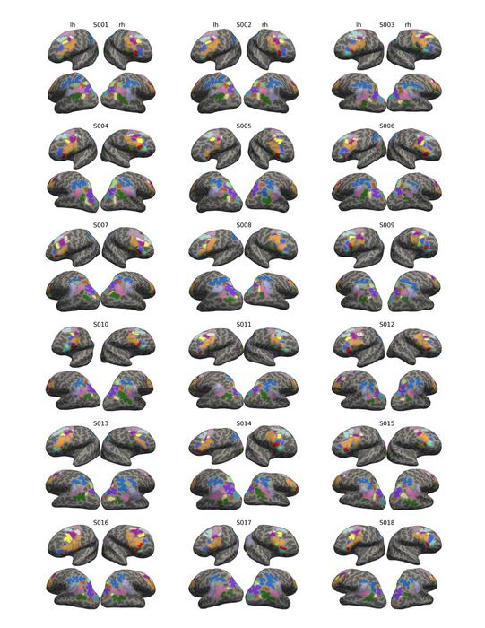
The Berkeley study enrolled 43 participants between the ages of 7 and 18, employing high-resolution magnetic resonance imaging (MRI) alongside functional magnetic resonance imaging (fMRI) to map both structural and functional features at an individual level. By focusing on 21 sulci per hemisphere—paying particular attention to the tertiary sulci in the lateral prefrontal and parietal cortices—the team meticulously quantified sulcal depth and assessed the strength of inter-regional functional connectivity. Participants also engaged in reasoning tasks during scanning, allowing researchers to directly link sulcal anatomy with cognitive performance and corresponding network activity.
Intriguingly, deeper tertiary sulci were consistently associated with higher “network centrality,” indicating that these grooves align with hubs of functional connectivity instrumental in reasoning processes. This relationship suggests that the geometry of brain folds is not an incidental outcome but may actively scaffold the underlying neural pathways, supporting complex cognitive operations. The findings challenge previous assumptions that cortical folding occurs haphazardly and instead point to sulcal morphology as a stable, individual-specific feature that could influence cognitive trajectory from childhood through adolescence.
The study’s senior authors, Professors Silvia Bunge and Kevin Weiner, highlighted the significance of examining sulcal morphology at the individual level. Historically, brain research has relied heavily on averaged brain atlases that obscure personal variations in sulcal patterns, often glossing over tertiary sulci that differ markedly among individuals. Weiner emphasized how these inter-individual variations in tertiary sulci have long posed a conundrum, with his own investigative journey beginning as an undergraduate curiosity about irregular cortical features that did not conform to standard brain maps. Their team’s pioneering work now opens the door for sulcal landmarks to serve as individualized anatomical references, revolutionizing how neuroscientists interpret brain function and structure.
Beyond establishing structural-functional correlations, this research frames tertiary sulci as potential biomarkers linking brain morphology with cognitive disorders. For example, earlier studies by the same lab identified the mid-fusiform sulcus—a tertiary sulcus implicated in face recognition—to be shorter and shallower in people with developmental prosopagnosia, a condition characterized by face blindness without evidence of brain damage. This precedent adds weight to the notion that tertiary sulcal variability is not just a benign anatomical feature but can be integral to understanding neurodevelopmental and cognitive differences.
The implications extend well beyond diagnostic markers. The study also touches on the plasticity inherent in sulcal depth and development. While the gross anatomical configuration of sulci is stable, their depth and cortical thickness can fluctuate with experience and development. Professor Bunge cautions against viewing sulcal features as deterministic, emphasizing instead the interplay between biology and environmental factors such as education, which profoundly shape individual cognitive trajectories. This nuance underscores that while sulcal morphology may set a neuroanatomical foundation, the ultimate expression of reasoning abilities is highly malleable and responsive to experiential inputs.
A notable technical advancement emerging from this work is the development of a computational tool designed to automate the identification of tertiary sulci in brain scans. Traditionally, neuroimaging software catalogs about 35 sulci, omitting many smaller or less prominent tertiary sulci. Weiner’s lab is expanding this repertoire to include over 100 sulci, leveraging machine learning to capture a more comprehensive map of cortical folding. This innovation promises to enhance reproducibility and precision across studies and enable clinicians and scientists alike to incorporate sulcal anatomy into broader neurodevelopmental assessments.
The study’s approach exemplifies a paradigm shift in neuroscience—moving away from generalized, averaged brain models to prioritize individual anatomical variation. By anchoring measures of functional connectivity to personalized sulcal morphology, the research navigates the complexities introduced by inter-individual differences that have historically hindered the interpretation of brain-behavior relationships. This fine-grained anatomical focus facilitates a more accurate understanding of how the local structure of the brain connects to network-level function and ultimately cognitive performance.
From an evolutionary perspective, the elaboration of tertiary sulci correlates with areas of the cortex that have undergone significant expansion in humans, especially those linked with advanced cognitive capabilities like reasoning, planning, and self-control. This pattern further suggests that tertiary sulci may represent anatomical adaptations supporting the cognitive versatility distinctive to Homo sapiens. Their protracted development, continuing into adolescence, dovetails with the extended maturation of executive functions, underscoring the developmental logic of sulcal formation alongside cognitive milestones.
The researchers acknowledge that while deeper tertiary sulci correlate with more efficient neural connectivity and better reasoning, the causal pathways remain to be fully elucidated. It is unclear whether sulcal depth directly enhances functional communication or is a surrogate marker for other microstructural features such as dendritic density, myelination, or synaptic architecture. Future studies employing multimodal neuroimaging and longitudinal designs are necessary to unravel these complex interrelations and to assess how sulcal anatomy interacts dynamically with cognitive development over time.
In conclusion, this seminal work reframes human brain folding as a purposeful and functionally relevant trait intricately tied to individual cognitive abilities. By spotlighting tertiary sulci—the brain’s subtle, last-developing creases—this research opens compelling avenues for understanding variability in reasoning skills among children and adolescents. It challenges entrenched neuroscience dogma, inviting a reconsideration of how cortical morphology shapes the neural networks that underpin human intelligence. As computational tools advance and datasets grow richer, the prospect of integrating sulcal anatomy into precision neuroscience and clinical practice becomes an exciting frontier.
Subject of Research: People
Article Title: Anchoring functional connectivity to individual sulcal morphology yields insights in a pediatric study of reasoning
News Publication Date: 19-May-2025
Web References: http://dx.doi.org/10.1523/JNEUROSCI.0726-24.2025
References: Hakkinen et al, UC Berkeley, as cited in The Journal of Neuroscience
Image Credits: Hakkinen et al, UC Berkeley
Keywords: tertiary sulci, brain folding, cortical morphology, functional connectivity, lateral prefrontal cortex, lateral parietal cortex, reasoning ability, neurodevelopment, brain imaging, pediatric neuroscience
Tags: biological factors in cognitive developmentbrain connectivity and reasoningcortical surface and cognitive variabilityevolutionary significance of sulcigroovy brain structuregroundbreaking neuroscience studiesimpacts of brain folds on efficiencylateral parietal cortex roleslateral prefrontal cortex functionsneural efficiency in children and adolescentssulci depth and brain communicationtertiary sulci and cognitive performance

