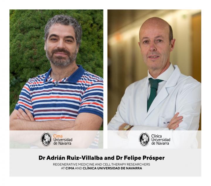
Cima and the Clinica Universidad de Navarra lead an international research showing the molecular mechanisms involved in the activation of these cardiac fibroblast in the presence of cardiac damage

Credit: Cima Universidad de Navarra
Pamplona (Spain), September 29th, 2020. Researchers at Cima and the Clinica Universidad de Navarra (Spain) have led an international study identifying the cardiac cells responsible for repairing the damage to this organ after infarction. These “restorative” cells are a subpopulation of cardiac fibroblasts that play a fundamental role in the creation of the collagen scar needed to avoid the rupture of the ventricular wall. The research also reveals the molecular mechanisms involved in the activation of these cells and the regulation of their function.
This finding, in which basic and clinical researchers have participated, will permit the identification of new therapeutic targets and the development of targeted therapies which will control the healing process of the heart after infarction.
The study has been published in the latest issue of the journal Circulation, the leading scientific journal of the American Heart Association.
Characterization of the reparative cardiac fibroblasts
Cardiac fibroblasts are one of the fundamental components of the heart. These cells play an essential role in maintaining the structure and mechanism of this vital organ. “Recent studies have shown that fibroblasts do not respond homogeneously to heart injury. Therefore the object of our study was to determine their heterogeneity during the remodeling of the injured ventricle and to understand the mechanisms that regulate the function of these cells”, said Dr Felipe Prósper, a researcher at Cima and the Clinica Universidad de Navarra, the leader of the study.
“Using single-cell transcription analysis techniques (single-cell RNA-seq), we identified a subpopulation within the cardiac fibroblasts, which we have named Reparative Cardiac Fibroblasts (RCF) due to their role after the cardiac injury. We have found that, when a patient has a heart attack, these RCF are activated and offer a fibrotic response due to which a collagen scar is generated to avoid the rupture of the cardiac tissue”, stated Dr Prósper, who is also a member of the Red de Terapia Celular (TerCel) and the Instituto de Investigación Sanitaria de Navarra (IdiSNA).
CTHRC1, a protein related to collagen and essential for the regenerative process
In the detailed molecular study, the researchers have found that the RCF have a unique transcriptional profile, that is to say, a specific information pattern for the expression of the genes involved in their cardiac function. “Among the main differential markers of the transcriptome of these cells, we have identified the CTHRC1 protein (Collagen Triple Helix Repeat Containing 1), a molecule with a fundamental role in the fibrotic response after myocardial infarction. Specifically, this protein participates in the collagen synthesis of the extracellular cardiac matrix and is crucial for the process of ventricular remodeling”, in the words of Adrián Ruiz-Villalba, a researcher on the Regenerative Medicine Program at Cima and first author of the article.
These results “suggest that the RCF activates the healing scar process of the cardiac lesion by secreting the CTHRC1 protein. Thus, this molecule may be considered as a biomarker associated with the physiological condition of the injured heart and a potential therapeutic target for patients who have suffered a heart attack or have dilated cardiomyopathy”, stated Ruiz-Villalba, who is also a researcher at IdiSNA. In addition to Cima and the Clinica Universidad de Navarra, basic and clinical researchers from the United States, Belgium and Austria have taken part in this research.
This work falls within the framework of the Cell Therapy and Regenerative Medicine research line being carried out at Cima and the Clinica Universidad de Navarra, aimed at understanding the regenerative potential of stem cells and their therapeutic application in different diseases such as cardiovascular ones. Specifically, this study is linked to the BRAV? project, an international research project combining bioengineering and cardiac stem cells to restore the function of an infarcted heart. BRAV? is an H2020 funded program by the European Union (H2020-SC1-BHC-07-2019-874827).
###
Bibliographical reference:
Ruiz-Villalba A. et al. Single-Cell RNA-seq Analysis Reveals a Crucial Role for Collagen Triple Helix Repeat Containing 1 (CTHRC1) Cardiac Fibroblasts after Myocardial Infarction. Circulation. 2020 Sep 25.
Doi: 10.1161/CIRCULATIONAHA.119.044557.
Media Contact
Miriam Salcedo de Prado
[email protected]
Original Source
https:/
Related Journal Article
http://dx.


