• HOME
  • NEWS
  • EXPLORE
    • CAREER
      • Companies
      • Jobs
    • EVENTS
    • iGEM
      • News
      • Team
    • PHOTOS
    • VIDEO
    • WIKI
  • BLOG
  • COMMUNITY
    • FACEBOOK
    • INSTAGRAM
    • TWITTER
Monday, August 18, 2025
BIOENGINEER.ORG
No Result
View All Result
  • Login
  • HOME
  • NEWS
  • EXPLORE
    • CAREER
      • Companies
      • Jobs
        • Lecturer
        • PhD Studentship
        • Postdoc
        • Research Assistant
    • EVENTS
    • iGEM
      • News
      • Team
    • PHOTOS
    • VIDEO
    • WIKI
  • BLOG
  • COMMUNITY
    • FACEBOOK
    • INSTAGRAM
    • TWITTER
No Result
View All Result
Bioengineer.org
No Result
View All Result
Home NEWS Science News Chemistry

Designer lens helps see the big picture

Bioengineer by Bioengineer
November 20, 2019
in Chemistry
Reading Time: 2 mins read
0
Share on FacebookShare on TwitterShare on LinkedinShare on RedditShare on Telegram

IMAGE

Credit: © 2019 KAUST


Microscopes have been at the center of many of the most important advances in biology for many centuries. Now, KAUST researchers have shown how a standard microscope can be adapted to provide even more information.

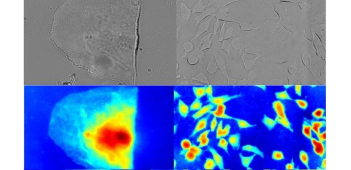
In its simplest form, microscopy creates an image of an object by measuring the intensity of light passing through it. This requires a sample that scatters and absorbs light in different ways. Many living cells, however, absorb very little visible light, meaning that there is only a small difference between light and dark regions, known as the contrast. This makes it difficult to see the finer detail.

But the light passing through the sample changes not only its intensity, but also its phase: the relative timing of the peaks in the optical wave. “Phase-contrast microscopy converts phase into larger amplitude variations and hence allows the viewing of fine, detailed transparent structures,” explains KAUST Ph.D. student Congli Wang.

Measuring the phase of light is trickier than measuring its intensity. Most phase-contrast microscopes must include a component that converts the phase change to a measurable intensity change. But this conversion is not precise; it only approximates the phase information.

Wang and his colleagues from the KAUST Visual Computing Center, under the supervision of Wolfgang Heidrich, a professor of computer science, have now developed a new method for quantitative phase and intensity imaging. Crucial to the performance of their microscope was an element known as a wavefront sensor. Wavefront sensors are custom-designed optical sensors that can encode the wavefront, or phase, information into intensity images.

The team designed an innovative high-resolution wavefront sensor, and the team members are now incorporating it into a commercial microscope to improve the performance of microscopy imaging. They then reconstructed the phase-contrast image using a computer algorithm they developed to numerically retrieved quantitative phase from an image pair: a calibration image obtained without the sample and a measurement image obtained with the sample in place.

This approach streamlines several aspects of microscopy. While other methods have achieved quantitative phase imaging in the past, they have required expensive or complicated setups, specialized light sources or a long time to generate the image. “Our method allows snapshot acquisition of high-resolution amplitude bright-field and accurate quantitative phase images via affordable simple optics, common white-light source and fast computations at video rates in real time,” says Heidrich. “It is the first time, to our knowledge, that all these advantages are combined into one technique.”

###

Media Contact
Carolyn Unck
[email protected]

Original Source

https://discovery.kaust.edu.sa/en/article/902/designer-lens-helps-see-the-big-picture

Related Journal Article

http://dx.doi.org/10.1038/s41598-019-50264-3

Tags: Algorithms/ModelsAudiovisual MediaComputer ScienceHardwareNanotechnology/MicromachinesSoftware EngineeringTechnology/Engineering/Computer Science
Share12Tweet8Share2ShareShareShare2

Related Posts

A Laser-Free Alternative to LASIK: Exploring New Vision Correction Methods

A Laser-Free Alternative to LASIK: Exploring New Vision Correction Methods

August 18, 2025
blank

Fe-Lattice O–O Ligands Boost Water Oxidation Catalysis

August 18, 2025

Innovative Technology Developed to Precisely Control Pore Wall Crystallinity

August 18, 2025

Future Reactors May Harness Nuclear Waste as a Fuel Source

August 18, 2025
Please login to join discussion

POPULAR NEWS

  • blank

    Molecules in Focus: Capturing the Timeless Dance of Particles

    141 shares
    Share 56 Tweet 35
  • Neuropsychiatric Risks Linked to COVID-19 Revealed

    80 shares
    Share 32 Tweet 20
  • Modified DASH Diet Reduces Blood Sugar Levels in Adults with Type 2 Diabetes, Clinical Trial Finds

    59 shares
    Share 24 Tweet 15
  • Predicting Colorectal Cancer Using Lifestyle Factors

    47 shares
    Share 19 Tweet 12

About

We bring you the latest biotechnology news from best research centers and universities around the world. Check our website.

Follow us

Recent News

A Laser-Free Alternative to LASIK: Exploring New Vision Correction Methods

Novel Small Molecule Shows Promise in Mitigating Acetaminophen-Induced Liver Injury

Mapping Key Kinase Mutations in Oral Cancer

  • Contact Us

Bioengineer.org © Copyright 2023 All Rights Reserved.

Welcome Back!

Login to your account below

Forgotten Password?

Retrieve your password

Please enter your username or email address to reset your password.

Log In
No Result
View All Result
  • Homepages
    • Home Page 1
    • Home Page 2
  • News
  • National
  • Business
  • Health
  • Lifestyle
  • Science

Bioengineer.org © Copyright 2023 All Rights Reserved.