


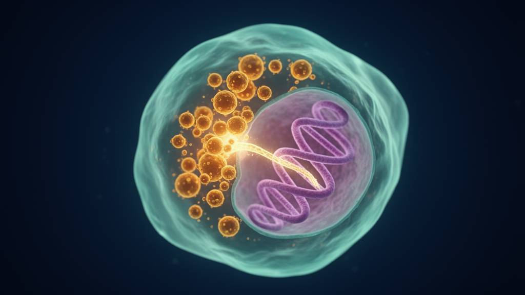
Dengue virus, an RNA virus with no native capacity to write itself into DNA, appears to do exactly that inside the cells of its mosquito vectors. A new study reports that after infection with dengue virus serotype 1 (DENV-1), two widely used Aedes mosquito cell lines produce detectable DNA copies of the viral genome, and that one of these cell lines yielded a chimeric sequence in which a fragment of viral DNA was joined to mosquito genomic DNA. The work, published in the journal Discover Viruses, also documents virus-derived DNA in wild-caught Aedes albopictus mosquitoes collected in Guangzhou, China, extending the observation beyond laboratory cultures into natural vector populations.
The research team, led by Xiang Guo, Qingqing Yin and colleagues with senior authorship by Xiaohong Zhou, set out to characterise a phenomenon that has been glimpsed before but never systematically mapped for dengue. DNA forms of the genomes of several RNA viruses, including Zika, West Nile, dengue, Chikungunya and La Crosse viruses, have previously been reported in mosquito cell cultures or in laboratory-reared mosquitoes. These fragments are thought to matter because they are closely associated with nonretroviral endogenous viral elements, abbreviated nrEVEs, which are stretches of viral sequence embedded in the genomes of a wide range of animals. In Aedes mosquitoes, such elements are believed to feed a heritable, nucleic-acid-based antiviral silencing system that acts through piRNAs, small RNAs that guide the immune response against future infections.
To follow the formation of viral DNA over time, the researchers established in vitro infection systems using two cell lines: C6/36 cells, derived from Aedes albopictus, and Aag2 cells, derived from Aedes aegypti. They infected the cells with the DENV-1 strain N46V and harvested cells and supernatant at 1, 3, 5, 7, 9 and 11 days after infection. After extracting DNA and treating it with DNase I, an enzyme that digests free DNA, they amplified viral sequences by PCR using 19 primer pairs spread across the dengue genome. DNase treatment served as a critical control: because the viral genome is RNA, any PCR signal that survives DNase digestion of the sample must originate from DNA rather than residual viral RNA carried over during extraction.
The timing of viral DNA appearance differed between the two cell lines. In C6/36 cells, viral DNA was first detected at 3 days post-infection, whereas in Aag2 cells it did not appear until 5 days post-infection. Once initiated, DNA synthesis persisted in both cell types through at least 11 days post-infection, the latest time point examined. Four additional DENV-1 strains isolated from patients during dengue outbreaks in Guangdong Province between 2014 and 2019, named ZS11, ZS23, ZS50 and 18488, showed the same pattern in C6/36 cells, with DNA forms appearing at 3 days post-infection, indicating that the phenomenon is not peculiar to a single viral isolate.
The coverage of the viral genome represented in DNA form was not uniform. Not every primer pair produced a band of the expected size, and statistical comparison showed that the genome coverage of viral DNA generated in C6/36 cells was significantly greater than in Aag2 cells, with a Wilcoxon test giving p < 0.05. Nevertheless, viral DNA arose from both the structural and the nonstructural regions of the DENV-1 genome. Using combined primer pairs, the team also amplified long fragments exceeding 1500 base pairs, which appeared concentrated in the C, E, NS2a, NS3, NS4a, NS4b and NS5 regions. This patchy but broad distribution echoes earlier findings that viral DNA forms in infected mosquitoes are fragmentary and do not span the entire viral genome.
The field component of the study drew on a monitoring programme in Guangzhou. From March 2015 to February 2017, the researchers had access to 3326 female and 711 male adult Aedes albopictus mosquitoes plus 6372 larvae, collected from six districts of the city. Pools of 10 to 15 individuals were screened for dengue RNA by nested RT-PCR and quantitative RT-PCR, and 30 of 717 pools tested positive for both. Of these, 24 pools remained available and were analysed for viral DNA. Using primers targeting the junction between the NS5 gene and the 3′ untranslated region of the viral genome, the team detected viral DNA fragments in 8 of the 24 pools, or 33.3 percent, and DNase digestion confirmed that the products derived from DNA rather than leftover RNA.
Sequencing of the field-derived viral DNA fragments revealed that they belonged to two separate clades of DENV-1 that had circulated in Guangzhou in overlapping years. One group of sequences, found in four pools, matched clade 1K1, which circulated during 2013 to 2015; the other, found in five pools, matched clade 5C1, which circulated from 2013 to 2019. Compared with a reference genome, some sequences shared a single nucleotide mutation, A10084G, together with a 21-base-pair deletion at the start of the 3′ untranslated region, while others resembled a different reference strain and shared a distinct mutation, A10295T. In one case, two variant sequences from the same clade came from a single pool, and in another, sequences from two different clades were recovered from the same pool, which the authors attribute to mixed mosquitoes within a sampling pool.
The central question was whether any of this viral DNA becomes integrated into the mosquito genome. To address it, the researchers constructed genome walking libraries from infected cells, digesting genomic DNA with four restriction enzymes, DraI, EcoRV, PvuII and StuI, ligating synthetic adaptors to the ends, and then amplifying outward from five viral regions using nested primers, with high-throughput sequencing of the products. From C6/36 cells infected with DENV-1, six walking fragments were obtained. One of them was a genuine host-virus chimera: a 528-base-pair DNA fragment from the C-prM region of the DENV-1 genome fused to a 482-base-pair sequence matching a contig of the Aedes albopictus genome. Adaptor and gene-specific primer sequences flanked the chimeric junction, supporting its authenticity as an amplification product rather than an artefact of alignment. The other five fragments aligned only to the viral genome, not to the mosquito genome. In Aag2 cells, tested with the same primer pairs, no comparable chimeric event was detected.
The authors interpret these results with appropriate caution. Viral DNA fragments were found at varying frequencies across samples, but the majority are likely nonintegrated, episomal DNA molecules rather than sequences stably inserted into chromosomes. Only a single host-virus chimeric event emerged from the screen, which the team reads as evidence that the probability of actual DENV-1 integration into the mosquito genome is low. They also note that four of the six walking products, including the chimeric flank, contained sequences matching the restriction sites used to build the libraries, a detail relevant to assessing how the observed junctions arise. The broader context is that while retroviruses carry their own reverse transcriptase and integrase, integration of nonretroviral RNA viruses has long been considered improbable; studies of lymphocytic choriomeningitis virus and Borna disease virus showed that host retroelements such as LINE-1 can mediate such integration, and the authors suggest an analogous mechanism may operate here, though this remains a hypothesis rather than a demonstrated pathway.
The study also situates its findings within the known landscape of viral fossils in Aedes genomes. Dozens of nonretroviral endogenous viral elements from several virus families have been found in the genomes of Aedes aegypti and Aedes albopictus, and a comparative analysis of the so-called EVEome found that 72 percent of nonretroviral integrated RNA virus sequences in Aedes species are homologous to Rhabdoviridae, 27 percent are close to the flaviviruses, and the remaining 1 percent belong to bunyaviruses and reoviruses. Against that backdrop, the demonstration that DENV-1 DNA forms arise within days of infection, persist for at least 11 days, occur in wild mosquito populations, and can at least occasionally join host genomic sequence, offers a plausible route by which new viral elements might enter the vector germline. The authors emphasise that further evaluation is needed to determine how often integration actually occurs, and that the biological consequences, whether viral DNA primes protective antiviral immunity or serves as raw material for viral evolution and host-pathogen coadaptation, remain open questions for future work in natural mosquito populations.
Subject of Research: Formation of dengue virus serotype 1 DNA fragments and a host-virus chimeric integration event in Aedes mosquito cells and wild-caught mosquitoes
Article Title: Evidence for virus-derived DNA and host–virus chimeric events generating in mosquito cell cultures following infection with dengue virus serotype 1
Article References: Guo, X., Yin, Q., Teng, P., Liu, X., Li, Z., Wang, Y., Xie, T., Hu, M., Ren, W., Liu, Y., Zhang, X., Chen, X.-G., & Zhou, X. (2026). Evidence for virus-derived DNA and host–virus chimeric events generating in mosquito cell cultures following infection with dengue virus serotype 1. Discover Viruses, 3(1), Article 12. https://doi.org/10.1007/s44370-026-00049-w
Image Credits: AI Generated
DOI: 10.1007/s44370-026-00049-w
Keywords: dengue virus, DENV-1, viral DNA, Aedes albopictus, Aedes aegypti, endogenous viral elements, genome integration, mosquito cells, piRNAs, Guangzhou, genome walking, arbovirus persistence
News Source: Juliet Wilcox. (October 29, 2026). Dengue virus leaves DNA traces and a rare genomic integration in mosquito cells. Scienmag.



