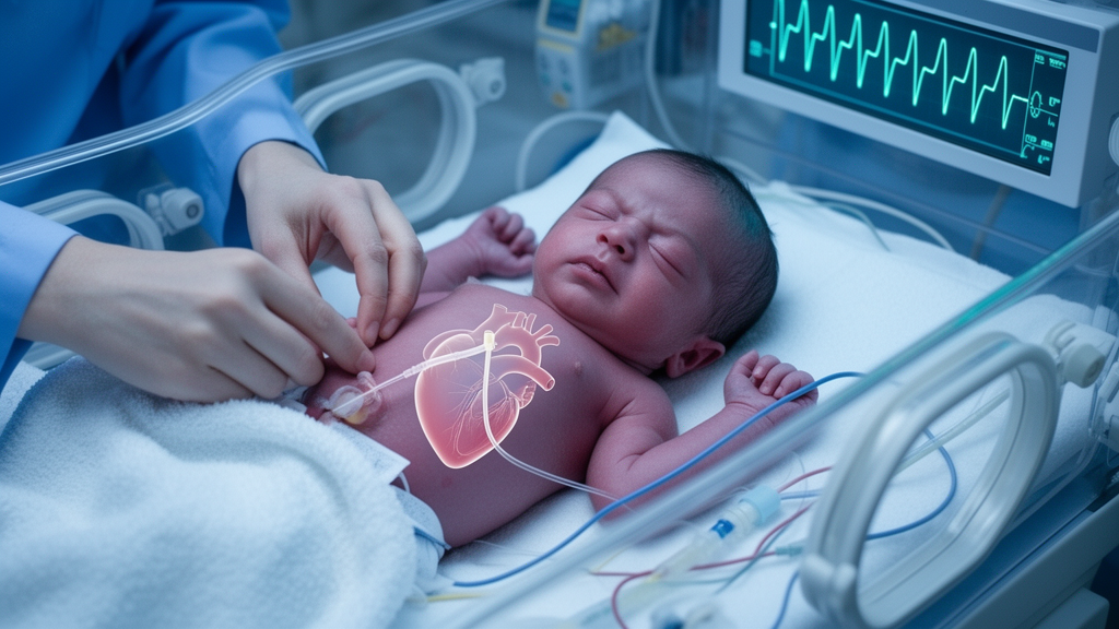
A 33-week-old neonate weighing 1,310 grams developed sudden respiratory deterioration on day 6 of life after initially improving with nasal continuous positive airway pressure, according to a BMC Pediatrics case report. Imaging identified a massive right pleural effusion, and subsequent investigation linked the deterioration to delayed migration of an umbilical venous catheter, with infused fluid entering the pleural space.
Umbilical venous catheters are often used in neonatal intensive care units and are generally considered relatively safe. Pleural effusion caused by extravasation of fluid from a catheter is an uncommon but potentially life-threatening complication. In the reported case, the catheter appeared appropriately positioned on the initial radiograph. Later imaging showed that its tip had migrated downward and developed a distal kink below the diaphragm, demonstrating that its position had changed after the earlier examination.
The infant’s abrupt respiratory decline required invasive ventilation. Chest radiography and bedside lung ultrasound showed a massive right pleural effusion. Clinicians drained the pleural space and obtained clear, straw-colored fluid classified as transudative, but the fluid had a markedly elevated glucose concentration. Together with the imaging findings and the infant’s intravenous fluid therapy through the umbilical venous catheter, this result suggested that infused fluid had extravasated into the pleural cavity. The combination of pleural-fluid glucose measurement and bedside point-of-care ultrasonography helped clinicians rapidly diagnose the catheter-related complication.
Removal of the umbilical venous catheter was followed by rapid resolution of the pleural effusion. The neonate then recovered completely, and the effusion did not recur. The temporal sequence, together with the evidence of catheter migration and the composition of the drained fluid, supports the conclusion that delayed catheter displacement caused the fluid leak and acute respiratory deterioration. The case also shows why a catheter that was well positioned earlier cannot necessarily be assumed to remain correctly positioned throughout continued use.
As a single case report, the study documents one instance of delayed umbilical venous catheter migration in a preterm neonate. It does not establish how frequently this complication occurs or how many infants would experience rapid recovery after catheter removal. The authors nevertheless emphasize serial assessment of catheter position throughout the entire catheterization period, noting that it may facilitate earlier detection of migration and potential complications. In this patient, bedside ultrasound revealed the large effusion, while the unusually high glucose concentration in the drained pleural fluid pointed toward extravasated intravenous fluid rather than an unexplained accumulation of fluid alone.
Subject of Research: Delayed umbilical venous catheter migration causing pleural effusion and respiratory deterioration in a preterm neonate
Article Title: Delayed umbilical venous catheter migration causing acute respiratory deterioration in a preterm neonate: a case report
Article References: Sahoo, D. P., Krishnegowda, V. K., & Nanda, D. (2026). Delayed umbilical venous catheter migration causing acute respiratory deterioration in a preterm neonate: a case report. BMC Pediatrics. https://doi.org/10.1186/s12887-026-07583-y
Image Credits: AI Generated
DOI: 10.1186/s12887-026-07583-y
Keywords: umbilical venous catheter, preterm neonate, catheter migration, pleural effusion, fluid extravasation, respiratory deterioration, neonatal intensive care, bedside ultrasound, case report, Delayed, umbilical, venous
News Source: Ophelia Keating. (October 24, 2026). Delayed umbilical catheter migration linked to respiratory decline in preterm neonate. Scienmag.