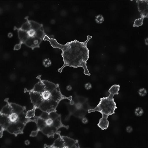
Influenza A virus (IAV) may target the lung’s most vulnerable cells not because they allow the virus to enter more efficiently, but because they are slower to mount an antiviral defense. A study published in the Chinese Medical Journal reports that alveolar type 2 (AT2) cells support substantially greater influenza replication than neighboring alveolar type 1 (AT1) cells. The difference appears to arise from a delayed and weaker innate immune response in AT2 cells, creating a window in which the virus can multiply before effective cellular defenses are activated. The finding offers a new explanation for how influenza infection damages the alveolar epithelium, the delicate tissue responsible for exchanging oxygen and carbon dioxide.
IAV remains a major global respiratory threat, causing as many as 650,000 respiratory deaths each year, according to widely cited international estimates. In severe cases, infection can progress to viral pneumonia, acute respiratory distress syndrome, and long-lasting structural damage that may contribute to pulmonary fibrosis. The alveoli are central to this process because they form the terminal gas-exchange compartments of the lung. AT1 cells are extremely thin and cover most of the alveolar surface, allowing gases to pass efficiently between air and blood. AT2 cells occupy a smaller surface area but perform essential functions, including producing pulmonary surfactant, regenerating damaged alveolar epithelium, and coordinating aspects of the local immune response.
The researchers, led by Professors Bin Cao and Ivan Fan-Ngai Hung, used a combination of animal experiments and primary alveolar cell models to examine how the two epithelial cell types respond to influenza. In mice infected with the pandemic A/California/07/2009 H1N1 strain, the team used time-resolved immunofluorescence staining to track viral antigens in the lung. At three days after infection, viral material was detected almost exclusively in AT2 cells, while AT1 cells showed little evidence of infection. By seven days, the proportion of AT2 cells lining the alveoli had fallen sharply, indicating that these cells were particularly susceptible to virus-associated loss in the infected lung.
The pattern observed in the mice was reproduced in a newly established primary alveolar epithelial cell culture system. This is significant because conventional respiratory cell lines often fail to preserve the specialized characteristics of primary lung cells. By developing a model containing alveolar epithelial cells that retain features of AT1 and AT2 biology, the investigators were able to compare viral infection in a controlled setting. The experiments showed that AT2 cells accumulated higher levels of influenza viral gene segments and supported more extensive viral replication than AT1 cells. The results therefore point to an intrinsic difference in cellular antiviral control rather than an effect caused solely by the surrounding lung environment.
The researchers next examined whether the difference could be explained by the abundance of receptors or other molecules needed for viral entry. Influenza viruses attach to sialic-acid-containing structures on the surface of respiratory cells before entering them through endocytosis. However, the study found no decisive receptor-abundance difference that could account for the preferential infection of AT2 cells. Instead, the major distinction emerged after infection had begun. Transcriptomic analysis showed that AT1 cells activated antiviral gene programs more rapidly and more strongly than AT2 cells, despite AT2 cells containing more viral genetic material.
This early immune response depends heavily on interferons, signaling proteins that alert infected and neighboring cells to the presence of a virus. Type I interferons, including interferon-alpha and interferon-beta, and type III interferons, particularly the interferon-lambda family, activate intracellular pathways that induce interferon-stimulated genes. These genes can interfere with multiple stages of the viral life cycle, including genome replication, protein synthesis, assembly, and release. The study found stronger induction of genes associated with type I and type III interferon signaling in AT1 cells. AT1 cells also showed more rapid activation of interferon-regulatory pathways, interferon-stimulated genes, cytokines, and chemokines, all of which can help restrict viral spread and recruit immune cells.
In AT2 cells, by contrast, these protective responses were delayed and attenuated. Quantitative polymerase chain reaction experiments confirmed the transcriptomic results, demonstrating lower or slower induction of key antiviral genes in the AT2 population. This creates a potentially important biological imbalance: AT2 cells may become heavily infected before they have produced enough interferons and antiviral proteins to limit replication. Once viral burden rises, the resulting cellular stress, inflammatory signaling, and cell death may contribute to disruption of the alveolar barrier. Loss of AT2 cells could be especially damaging because these cells help maintain surfactant production and serve as progenitors that replenish injured alveolar epithelium.
The findings challenge the assumption that AT1 cells should be influenza’s primary target simply because they cover most of the alveolar surface. Surface area alone does not determine susceptibility. The study suggests that the timing and strength of intracellular antiviral signaling can be more influential than the number of potential entry sites. It also distinguishes influenza injury from other forms of alveolar damage. For example, hyperoxia, or exposure to abnormally high oxygen concentrations, is known to cause prominent injury to AT1 cells. Influenza therefore appears to produce a different pattern of epithelial damage, one shaped by selective viral replication and cell-specific immune programming.
The results may have implications for antiviral development and for understanding why some respiratory infections progress from an upper-airway illness to severe disease in the deep lung. Drugs that directly inhibit influenza replication remain essential, but interventions that improve antiviral readiness in AT2 cells could eventually offer an additional strategy. Such approaches would need to be carefully controlled, because excessive interferon or inflammatory signaling can itself damage lung tissue. The primary alveolar models described in the study could help researchers test antiviral compounds, interferon-based treatments, and host-directed therapies while comparing their effects on AT1 and AT2 cells. They may also provide a platform for investigating how age, pre-existing lung disease, or prior immunity alters cell-specific responses.
The investigators emphasize that further work is needed before the conclusions can be translated directly to human disease. The present findings were obtained using mice, primary cell cultures, and the A/California/07/2009 H1N1 strain. Human alveolar cells may differ in their baseline antiviral programs, and highly pathogenic avian influenza viruses or other seasonal and pandemic strains may interact with AT1 and AT2 cells in different ways. Nevertheless, the study identifies a clear mechanism by which delayed innate immunity can increase susceptibility to influenza: AT2 cells do not necessarily permit more infection because they offer more entry receptors, but because their antiviral alarm system responds too slowly. By placing cell-specific immune timing at the center of alveolar injury, the work provides a new framework for understanding how influenza gains a foothold in the gas-exchange region of the lung.
Subject of Research: Cells
Article Title: Delayed Antiviral Immune Response in Alveolar Type 2 Cells Increases Susceptibility to Influenza Virus in Alveoli
News Publication Date: 10-Aug-2026
Web References: https://doi.org/10.1097/CM9.0000000000004107
References: Delayed Antiviral Immune Response in Alveolar Type 2 Cells Increases Susceptibility to Influenza Virus in Alveoli, Chinese Medical Journal, DOI: 10.1097/CM9.0000000000004107
Image Credits: Chinese Medical Journal
Keywords: Influenza A virus, H1N1, alveolar type 1 cells, alveolar type 2 cells, interferon, interferon-stimulated genes, antiviral immunity, lung infection, alveolar injury, respiratory disease
Tags: alveolar cell types in influenza infectionalveolar epithelium damage in influenzaalveolar type 2 cells immune responsealveoli structure and function in viral infectionsdelayed antiviral defense in lung cellsimpact of delayed immune response on influenza severityinfluenza A virus replication in alveolar cellsinfluenza susceptibilityinnate immune response in alveolar cellslung immune response timingpulmonary fibrosis and influenzarespiratory infections and lung cell vulnerability