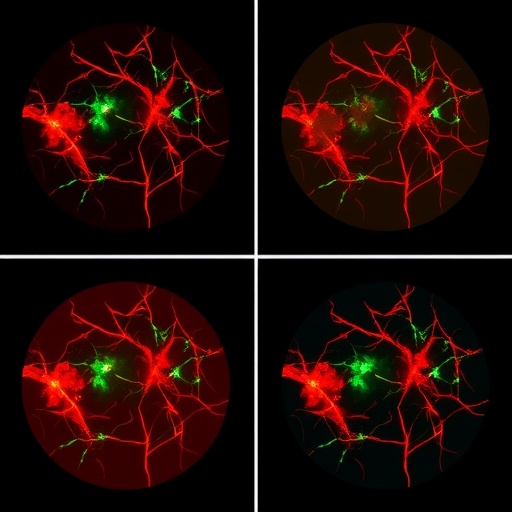
Researchers in China have unveiled a deep learning framework that can trace the finest branches of the blood vessel network inside the human eye with unprecedented reliability, offering a potential new tool for catching blinding diseases before irreversible damage occurs. The method, described in the Journal of Ambient Intelligence and Humanized Computing, combines a classical mathematical technique known as the stationary wavelet transform with a purpose-built multiscale neural network, and it has been shown to outperform existing approaches across three separate datasets of retinal images.
The retina, the thin layer of light-sensitive tissue lining the back of the eye, is the only place in the human body where blood vessels can be observed directly and non-invasively. This unique window makes retinal vessel imaging enormously valuable clinically. Changes in the width, curvature, branching pattern, and density of these vessels are early fingerprints of systemic and ocular conditions, including diabetic retinopathy, hypertensive retinopathy, and glaucoma, all of which can progress silently for years before a patient notices any loss of vision. Because early detection is the single most effective defense against permanent visual impairment, ophthalmologists have long sought automated systems capable of measuring the vasculature accurately and consistently. The manual tracing of vessels, however, is tedious, time-consuming, and subject to significant variability between different human experts, a problem the research team set out to eliminate.
The new study, led by Guanghui Song of Ningbo Tech University together with Binhua He of Zhejiang Sci-Tech University and Yan Nie of Ningbo University, focuses on a particularly challenging type of retinal imaging: fluorescein angiography, or FA. In this procedure, a fluorescent dye is injected into the bloodstream, and as it circulates through the retina, a specialized camera captures sequences of images in which the vessels glow brightly against a dark background. FA offers a dynamic, high-contrast view of blood flow that standard color fundus photography cannot match, revealing leakage, non-perfusion areas, and vascular abnormalities that would otherwise remain hidden. Yet the very characteristics that make FA so informative also make it difficult for automated analysis. Images are often affected by uneven illumination, background fluorescence, noise, and wide variation in vessel calibers, from thick major arcades down to capillaries only a pixel or two across.
To cope with this complexity, the researchers turned to multiscale analysis, a strategy rooted in the mathematics of wavelets. Wavelet transforms decompose a signal or image into components at different scales and resolutions, much as a musical equalizer separates sound into bass, midrange, and treble. The stationary wavelet transform in particular is translation-invariant, meaning it does not shift its representation when the input shifts, a property that makes it well suited to detecting structures of different sizes without introducing artifacts. By applying this transform to the angiographic images, the team generated a set of feature maps that emphasized vessels at each characteristic width. Fine capillaries standing out at fine scales, while large vessels emerged at coarser ones. This preprocessing step effectively converts the raw image into a richer representation in which vessel-like structures are amplified and background clutter suppressed.
These multiscale features then feed into a fully complex multiscale neural network, an architecture designed so that its internal processing parallels the scale hierarchy established by the wavelet stage. The core design principle is adaptation to two sources of variability that plague retinal image analysis: the enormous range of vessel widths and the constantly changing orientation of vessels as they sweep across the curved surface of the retina. Because the network receives explicit scale-decomposed inputs, it can assign different weights to different scales depending on context, learning to interpret a faint thin structure as a capillary rather than noise, and to distinguish a thick vessel from an illumination artifact. The complex-valued formulation of the network adds another dimension of representational power, allowing phase information carried by the wavelet coefficients to be exploited rather than discarded, which helps the model respond to the directionality of vascular structures.
Training such a network on relatively limited sets of expert-annotated medical images is a notorious bottleneck in medical artificial intelligence. Overfitting, in which a model memorizes the quirks of its training data rather than learning generalizable patterns, is a constant danger. The team addressed this with a deliberate data augmentation strategy: rotation operations were applied at least once across the layers during the training phase. In effect, the network was repeatedly confronted with the same vascular anatomy presented at different orientations, forcing it to learn features that are genuinely rotation-tolerant rather than tied to the particular angle at which vessels happened to appear in the training images. This echoes a broader theme in modern deep learning research, where equivariance to geometric transformations is prized precisely because biological structures, like retinal vessels, can appear at arbitrary orientations in any given scan.
The performance of the resulting system was evaluated on three different datasets, and in each case the proposed method delivered better results than the current state-of-the-art techniques it was compared against. This cross-dataset consistency matters greatly, because many published segmentation algorithms perform impressively on the specific dataset they were tuned to but degrade sharply when confronted with images from a different camera, a different patient population, or a different imaging protocol. Robustness across datasets is therefore one of the most honest indicators of whether a method has genuine clinical potential or is merely exploiting statistical idiosyncrasies of one benchmark.
Equally significant is the stability of the method. The researchers report that the framework produces consistent results across different training datasets and across inter-rater variability, the well-documented phenomenon in which two human experts annotating the same image disagree on fine details, particularly around the thinnest vessels and at branch points. A diagnostic tool that fluctuates depending on which annotator labeled its training data is of limited clinical value. The ability of the new method to absorb and transcend this human disagreement suggests it has learned a representation of vascular anatomy that is closer to the underlying biological reality than to the idiosyncrasies of any single annotation. As the authors note, this means the method can be practically used anywhere, a claim of portability that, if validated in prospective clinical studies, would be a meaningful advance for screening programs in settings where expert graders are scarce.
The clinical implications extend well beyond the technical achievement of drawing cleaner vessel maps. In diseases such as diabetic retinopathy, which affects a substantial fraction of the world’s growing diabetic population, the earliest signs of pathology are vascular: microaneurysms, capillary dropout, and changes in vessel tortuosity. Automated and accurate vessel segmentation is the foundational step upon which all such quantitative measurements depend. Fluorescein angiography is particularly central to assessing retinal perfusion, and recent work from other groups has explored using deep learning even to synthesize FA-like information from color fundus photographs. The present study complements that line of research by tackling the segmentation problem directly on authentic angiographic images, where the fluorescent signal provides a rich but noisy substrate for machine analysis.
The methodology also illustrates a persuasive middle path in medical AI design. Rather than relying solely on a generic convolutional architecture and hoping that enough data will teach it everything, the team built explicit domain knowledge into the pipeline. The choice of the stationary wavelet transform encodes decades of signal-processing understanding about how structures of varying scale can be separated, and the multiscale network architecture mirrors the physical fact that retinal vessels span a wide range of calibers. This hybrid of classical mathematics and modern deep learning, sometimes described as physics-informed or knowledge-guided machine learning, is increasingly seen as the most promising route to systems that are accurate, data-efficient, and trustworthy, the three qualities that regulators and clinicians alike demand from medical technology.
The work, which was partially supported by the Ningbo “Science and Technology Innovation Yongjiang 2035” key technology breakthrough plan project, arrives at a moment when the burden of retinal disease is rising worldwide. Diabetic retinopathy alone remains a leading cause of preventable blindness among working-age adults, and hypertension-related vascular changes in the retina are increasingly recognized as markers of broader cardiovascular risk. Screening programs that could deploy a stable, dataset-agnostic segmentation algorithm on angiographic images would multiply the reach of limited ophthalmological expertise, flagging patients who need urgent attention while sparing healthy individuals unnecessary interventions.
The authors caution, as with any new method, that broader clinical validation will be needed before deployment in routine care, and the study’s three-dataset evaluation, while encouraging, represents a step on a longer road toward regulatory approval and integration into hospital workflows. Nevertheless, the combination of superior performance, robustness across datasets, and stability against human annotation variability marks this framework as a notable contribution to the rapidly evolving field of retinal image analysis. If the promise holds, the humble wavelet, a mathematical tool first formalized more than a century ago in the work on orthogonal function systems, may find itself at the heart of software that safeguards the eyesight of millions.
Subject of Research: Deep learning-based segmentation of retinal blood vessels in fluorescein angiography images, combining stationary wavelet transform multiscale analysis with a multiscale neural network for diagnosing retinal diseases.
Subject of Research: Technology and Engineering
Article Title: Deep learning-based segmentation of retinal blood vessels in fluorescein angiography
Article References: Song, G., He, B., & Nie, Y. (2026). Deep learning-based segmentation of retinal blood vessels in fluorescein angiography. Journal of Ambient Intelligence and Humanized Computing, 17(5), 1371-1383. https://doi.org/10.1007/s12652-026-05104-4
Image Credits: AI Generated
DOI: 10.1007/s12652-026-05104-4
Keywords: retinal blood vessels, segmentation, deep learning, fluorescein angiography, stationary wavelet transform, multiscale neural network, diabetic retinopathy, retinal disease diagnosis, fundus imaging, medical image analysis, computer vision, ophthalmology
Cite Scienmag News
APA MLA Chicago
Blake Davidson. (September 4, 2026). Deep learning segments retinal blood vessels in fluorescein angiography images. Scienmag. https://scienmag.com/deep-learning-segments-retinal-blood-vessels-in-fluorescein-angiography-images/
Blake Davidson. “Deep learning segments retinal blood vessels in fluorescein angiography images.” Scienmag, 4 September 2026, https://scienmag.com/deep-learning-segments-retinal-blood-vessels-in-fluorescein-angiography-images/. Accessed 4 September 2026.
Blake Davidson. “Deep learning segments retinal blood vessels in fluorescein angiography images.” Scienmag. September 4, 2026. https://scienmag.com/deep-learning-segments-retinal-blood-vessels-in-fluorescein-angiography-images/
Copy citation Download RIS
Tags: advanced image processing for eye healthAI-based glaucoma detection methodsautomated detection of diabetic retinopathyautomated retinal vessel detectionblood vessel network analysis in the human eyecomputer-aided diagnosis for glaucomadeep learning applications in ophthalmologydeep learning for early ocular disease diagnosisdeep learning in ophthalmologydeep learning retinal blood vessel segmentationearly diagnosis of diabetic retinopathyfluorescein angiography image analysisimage processing in eye health monitoringmultiscale neural network for retinal imagingmultiscale neural network for retinal vesselsnon-invasive eye imaging techniquesnon-invasive retinal vessel imaging techniquesretinal blood vessel segmentationretinal blood vessel segmentation datasetsretinal image dataset analysisstationary wavelet transform in medical imagingvessel morphology analysis in retinal images