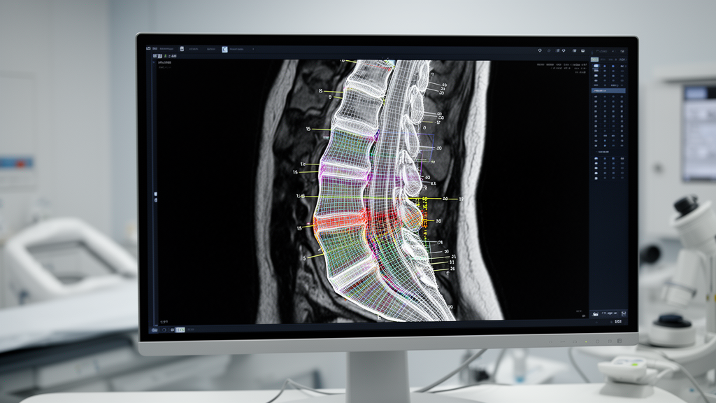
A deep learning pipeline integrating oriented object detection and transformer-based semantic segmentation has achieved submillimeter measurement agreement with senior radiologists when assessing lumbar vertebral morphology on sagittal T1-weighted magnetic resonance imaging. Developed by a multidisciplinary research team at the Department of Radio-Diagnosis within SRM Medical College Hospital and Research Centre, the automated methodology demonstrated mean absolute measurement errors of 0.640 millimeters for anterior vertebral height, 0.691 millimeters for central height, and 0.612 millimeters for posterior height across an evaluation cohort comprising 1,122 vertebral levels from 190 patient examinations. When quantifying maximum vertebral body height loss across wedge, biconcave, and crush deformity patterns, the automated system recorded an overall mean absolute error of 2.51 percentage points compared with reader-derived reference values, with 88.1 percent of all automated deformity calculations falling within five percentage points of expert assessments. Rather than operating as an isolated image classification model, the computational workflow functions as an end-to-end framework that takes raw magnetic resonance DICOM series as input, performs automated mid-sagittal slice selection, extracts physical calibration data, localizes tilted vertebral bodies, segments anatomical margins, and generates reviewable graphical measurement overlays alongside tabulated morphometric indices.
Low back pain and degenerative lumbar spine disorders represent leading contributors to global disability and healthcare expenditure, necessitating frequent radiological evaluation in routine clinical practice to investigate disc degeneration, neural element compression, spinal canal stenosis, spondylolisthesis, and structural bone abnormalities. Within this clinical setting, reduction in vertebral body height serves as a fundamental radiological indicator reflecting osteoporotic compression fractures, acute traumatic structural collapse, or chronic biomechanical weakening. Historically, quantitative vertebral morphometry has been conducted on lateral projection spine radiographs by placing manual caliper lines across the anterior, central, and posterior margins of vertebral bodies to define wedge, biconcave, and crush deformities. While magnetic resonance imaging is primarily acquired to evaluate soft-tissue contrast, neural compromise, and bone marrow signal alterations rather than serving as the historical primary tool for geometric morphometry, sagittal lumbar MRI sequences intrinsically contain rich anatomical structural data. However, manual morphometric evaluation directly on clinical PACS workstations remains labor-intensive, time-consuming, and susceptible to substantial interobserver variability arising from physiological lumbar lordosis, individual patient positioning, degenerate endplate irregularities, marginal osteophytes, and subjective landmark placement by human readers.
To establish a reproducible and objective computational workflow capable of overcoming these operational challenges, the researchers designed a retrospective methodological investigation spanning November 2025 through March 2026 utilizing de-identified, publicly accessible spine datasets without prospective patient enrollment. The internal development and testing cohort was derived from the RSNA Lumbar Degenerative Imaging Spine Classification dataset, which initially contained 1,975 lumbar MRI examinations. A Python script randomly sampled 1,000 cases without replacement or preselection for specific spinal abnormalities, which were subsequently screened for sagittal T1-weighted image quality. After excluding 90 technically degraded cases characterized by severe noise, insufficient contrast, or obscured vertebral boundaries, 910 internal cases were retained and divided into 729 training examinations, 91 validation examinations, and 90 internal test examinations containing 549 test vertebrae. To test model generalization against unseen external imaging sources, 100 cases encompassing 573 vertebral levels were randomly sampled without replacement from an independent Mendeley lumbar spine MRI repository containing 515 examinations. Combined, the final morphometric testing cohort encompassed 190 independent cases and 1,122 distinct vertebral levels, providing a comprehensive validation benchmark across diverse patient presentations.
Establishing rigorous ground-truth reference standards required extensive manual annotation conducted by two senior radiologists, each possessing more than ten years of specialized experience in clinical spine MRI interpretation. The imaging examinations were partitioned equally between the two primary readers, with each case annotated once, followed by independent adjudication and anatomical verification by a third senior radiologist with fourteen years of specialized spine imaging experience. To train the automated mid-sagittal slice-selection model, internal sagittal DICOM series comprising 13,943 slices across 820 training and validation examinations were converted into NIfTI format and opened in ITK-SNAP to manually delineate binary spinal canal segmentation masks. For vertebral region-of-interest localization and segmentation, representative sagittal images were imported into the CVAT platform, where vertebral bodies were manually outlined with closed polygon contours and subsequently transformed into binary masks. Oriented bounding-box labels were subsequently generated by fitting minimum-area rotated rectangles around each mask with standardized margins, ensuring that localization boxes captured vertebral orientation without defining the anatomical boundary endpoints used for downstream physical height measurement.
The computational implementation was realized using Python 3.12 within Jupyter Notebook environments on a 64-bit Windows 11 workstation powered by an AMD Ryzen 7 7840HS processor, 16 gigabytes of system memory, and an NVIDIA GeForce RTX 4050 Laptop graphics processing unit equipped with six gigabytes of dedicated video memory. Region-of-interest localization was engineered utilizing the Ultralytics YOLOv8 framework, specifically deploying a YOLOv8n-OBB oriented bounding-box architecture trained for 100 epochs using the AdamW optimizer with an initial learning rate of 0.001, a batch size of eight, standard online augmentations, and automatic mixed precision. For mid-sagittal slice determination and fine vertebral segmentation, the team adapted pretrained SegFormer-B2 transformer networks from Hugging Face Transformers and PyTorch, which process overlapping image patch embeddings through four hierarchical transformer encoder stages alongside multilayer perceptron decoding blocks. A binary SegFormer-B2 model was trained at 512-by-512 pixel resolution for 40 epochs with a batch size of two to segment the spinal canal, whereas a separate two-class SegFormer-B2 network was trained at 256-by-256 resolution with a batch size of eight to delineate vertebral bodies from localized crops.
Inference execution within the deployed Tkinter desktop application begins when an operator selects a local folder containing unorganized DICOM files, prompting the software to automatically discover series metadata, extract protocol descriptions, and isolate the targeted sagittal T1-weighted acquisition. Image volumes are loaded using SimpleITK, normalized slice-wise to an eight-bit scale ranging from 0 to 255 while assigning zero intensity to degraded slices exhibiting intensity variations below 10⁻⁶, and converted to three-channel tensors. Crucially, the system extracts row and column pixel dimensions directly from DICOM tags 0028,0030 or 0018,1164, falling back to SimpleITK object metadata when necessary to ensure accurate physical scaling into millimeter units. To identify the optimal mid-sagittal plane, every sagittal slice is evaluated by the spinal canal SegFormer-B2 model, which outputs a probability map thresholded at 0.50 to calculate total cross-sectional canal area. The slice yielding the maximal predicted spinal canal area is designated as the primary mid-sagittal index, while candidate slices are stored and ranked to allow clinical users to manually inspect or select alternative adjacent slices whenever severe structural scoliosis or anatomical variations warrant human adjustment.
Once the primary mid-sagittal slice is established, the pipeline incorporates the immediately adjacent sagittal slices to form a three-slice evaluation set designed to mitigate single-plane measurement instability. Each slice is processed by the YOLOv8-OBB model at 1,024-pixel input resolution with a confidence threshold of 0.10 and a non-maximum suppression threshold of 0.70 to detect tilted vertebral bodies as four oriented corner coordinates. Using perspective transformation matrices derived from these corner coordinates, each detected vertebral body is extracted as an independent, rotation-normalized crop, neutralizing spinal curvature before semantic segmentation. The cropped image is resized to 256-by-256 pixels, segmented by the vertebral SegFormer-B2 model, and refined through post-processing that retains only the largest connected foreground component while executing morphological opening with a 3-by-3 elliptical structuring element. Cleaned segmentation masks are then mapped back into original DICOM coordinates via inverse perspective transformation. Vertebrae contacting the upper two image pixels are flagged and excluded from downstream morphometry to avoid erroneous calculations on truncated anatomical structures at image borders.
Automated anatomical labeling is conducted on the central reference slice by ordering detected vertebral centroids vertically from superior to inferior and applying a bottom-to-top sequence that identifies the lowest detected lumbar vertebra as L5 before sequentially naming L4, L3, L2, L1, and visible lower thoracic levels. To establish measurement boundaries within the aligned coordinate framework of each cleaned mask, anterior and posterior vertical zones are defined within side bands spanning 25 percent of the total mask width, connecting the superior-most and inferior-most mask boundary coordinates to define anterior and posterior height lines. The central height line is determined as the absolute minimum vertical pixel distance between upper and lower endplate boundaries within a central horizontal band, supported by a 60-pixel lateral search radius if central columns contain empty pixels. Endpoint coordinates are converted into physical millimeters using extracted row and column spacing. Detections on adjacent slices are matched to anchor center-slice vertebrae using vertical centroid proximity within a 60-pixel threshold, enabling three-slice median height aggregation across anterior, central, and posterior dimensions.
Using median-aggregated height values, the pipeline computes mean vertebral height alongside quantitative deformity percentages, calculating anterior wedge loss relative to posterior height and biconcave loss relative to the average of anterior and posterior dimensions. For crush deformity assessment, the algorithm calculates a reference baseline height derived from the 95th percentile of mean heights among preserved vertebrae exhibiting wedge and biconcave losses below an internal 18 percent threshold, mitigating the distorting influence of extreme individual anatomical dimensions. Maximum height loss is subsequently assigned as the largest percentage among wedge, biconcave, and crush parameters. Across the test cohorts, the automated mid-sagittal slice finder demonstrated Top-1 agreement with radiologists of 91.1 percent internally and 89.0 percent externally, with Top-3 accuracies reaching 100 percent and 99.0 percent. The YOLOv8-OBB detector achieved mAP50–95 scores of 0.760 internally and 0.730 externally, while SegFormer-B2 attained mean Dice similarity coefficients of 0.914 and 0.898, reflecting robust technical segmentation performance.
Statistical evaluation incorporating patient-level cluster bootstrapping across 5,000 resamples confirmed close concordance between automated measurements and consensus radiologist benchmarks, which themselves demonstrated excellent inter-reader reliability with intraclass correlation coefficients exceeding 0.95 across all height dimensions. Automated anterior, central, and posterior height measurements exhibited Pearson correlation coefficients of 0.957, 0.939, and 0.958 across the combined cohort, with Bland-Altman mean biases remaining minimal at 0.065 millimeters, -0.237 millimeters, and -0.194 millimeters. Stratified subgroup analyses revealed that while morphometric accuracy remained exceptionally high in nondeformed vertebrae, measurement error increased progressively with severe morphological collapse: mean absolute error for maximum height loss rose from 2.375 percentage points in grade 0 deformities to 4.450 percentage points in grade 3 deformities. The authors emphasize that the workflow is intended strictly as an objective, reviewable quantitative aid rather than an autonomous diagnostic system, noting that prospective multi-reader trials and validation across severe scoliosis, transitional anatomy, metallic implants, and diverse magnetic resonance field strengths will be required prior to routine clinical deployment.
Subject of Research: Automated lumbar vertebral morphometry using deep learning pipelines on sagittal T1-weighted magnetic resonance imaging
Article Title: An end-to-end pipeline using YOLOv8-OBB and SegFormer-B2 for automated lumbar vertebral morphometry on T1 sagittal MR images
Article References: Nixon, A., Lazar, V. R., Pendem, S., Rajendran, H. C., Aiyappan, S. K., Perumal, S. R., & Sekar, K. (2026). An end-to-end pipeline using YOLOv8-OBB and SegFormer-B2 for automated lumbar vertebral morphometry on T1 sagittal MR images. Discover Artificial Intelligence, 6(1), Article 1767. https://doi.org/10.1007/s44163-026-02503-6
Image Credits: AI Generated
DOI: 10.1007/s44163-026-02503-6
Keywords: vertebral morphometry, lumbar spine MRI, SegFormer-B2, YOLOv8-OBB, deep learning, vertebral compression, height loss measurement, oriented bounding boxes, spinal canal segmentation, medical image processing, DICOM analysis, transformer architecture, orthopedic imaging, radiological artificial intelligence, lumbar lordosis, interobserver agreement, computer-assisted radiology
News Source: Blake Davidson. (November 13, 2026). Deep Learning Pipeline Automates Lumbar Vertebral Morphometry on Sagittal Spine MRI. Scienmag.