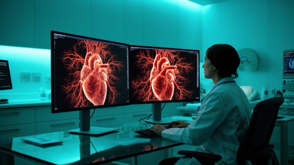
In cardiovascular imaging, photon-counting detector computed tomography marks a notable technological breakthrough over conventional CT systems, providing a threefold increase in spatial resolution alongside high temporal resolution. However, ultrahigh-resolution coronary computed tomography angiography scans are constrained by elevated image noise, which can compromise vessel-wall contour evaluation, obscure fine anatomic structures, and complicate luminal stenosis measurements. To address this technical trade-off, an exploratory study evaluated whether deep learning post-processing incorporating simultaneous noise suppression and iodine-specific contrast enhancement could improve image quality. Applying a vendor-agnostic deep learning tool to ultrahigh-resolution scans from twenty-five patients with coronary artery disease, researchers observed marked improvements in visual image quality, objective signal metrics, and vessel delineation across multiple reconstruction strengths. The findings indicate that deep learning algorithms effectively mitigate the noise penalty inherent to ultrahigh-resolution photon-counting scanning, delivering incremental technical benefits beyond standard vendor-specific iterative reconstruction algorithms.
The retrospective, single-centre study was conducted following formal approval from the local institutional Ethics Committee under protocol 2022-16359, complying with the 2008 Declaration of Helsinki. The study sample was derived from consecutive patients presenting with stable chest pain who were referred for coronary computed tomography angiography to exclude or evaluate coronary artery disease. Enrollment required ultrahigh-resolution photon-counting scanning based on an Agatston coronary artery calcium score threshold of ten or greater. Complete clinical datasets were acquired for all twenty-five enrolled individuals, comprising fifteen men and ten women with a mean age of 65.6 ± 10.6 years. An a priori power calculation in GPower 3.1 based on published effect sizes (dz of 2.08 for SNR, 2.11 for CNR, and 2.80 for slope) determined that five to six subjects were sufficient to achieve 0.95 power. Enrolling twenty-five participants ensured post hoc power exceeding 0.97 for all primary parameters under two-tailed testing with alpha set at 0.05.
Scans were acquired using a first-generation dual-source photon-counting detector CT system, the NAEOTOM Alpha from Siemens Healthineers, configured with two cadmium-telluride detectors offering 144 × 0.4 mm collimation. Scanning was performed in ultrahigh-resolution mode utilizing 120 × 0.2 mm collimation, a tube voltage of 120 kVp, and a gantry rotation time of 0.25 seconds. Patient preparation followed a standardized protocol: each participant received 0.8 mg of sublingual nitroglycerin approximately five minutes before acquisition to induce coronary vasodilation. When resting heart rates exceeded seventy beats per minute, intravenous metoprolol was administered up to 25 mg (mean ± standard deviation, 10.2 ± 7.0 mg). Electrocardiographic gating was selected according to cardiac rhythm: sequential scanning with prospective end-diastolic triggering for heart rates below seventy beats per minute, an extended prospective window for higher regular heart rates, and retrospective spiral electrocardiographic gating for patients exhibiting cardiac arrhythmias.
Intravascular contrast enhancement was managed using an initial test bolus procedure to determine circulation transit time, followed by a biphasic injection protocol. Patients received 70 mL of iopromide at an iodine concentration of 370 mg/mL (Ultravist 370) injected at a continuous rate of 5 mL/s, accompanied by an immediate 25-mL saline flush. Raw projection data were reconstructed at an ultra-thin slice thickness of 0.2 mm and a reconstruction increment of 0.1 mm using sharp vascular kernels designated as Bv64 on a dedicated off-site workstation. Scans were reconstructed without iterative filtering (Qoff), analogous to conventional filtered-back projection, and across all available strength levels of vendor-specific quantum iterative reconstruction spanning levels Q1, Q2, Q3, and Q4.
All reconstructed DICOM image volumes were subsequently processed with ClariACE, a vendor-agnostic deep learning post-processing software package developed by ClariPi. In this architecture, the network predicts iodine-component pixel intensities, amplifies them via a user-defined boosting factor, and combines the scaled representation with the original input image while applying denoising operations. The tool provides three adjustable parameters—denoising strength, edge bending, and contrast enhancement—each configurable across a continuous scale ranging from zero (no processing) to one (maximum modification). To determine the optimal operational configuration for coronary anatomical depiction, two experienced radiologists possessing five and seven years of clinical imaging experience systematically tested multiple setting combinations. Following joint visual review, the readers established that the maximum setting combination—denoising strength 1, edge bending 1, and contrast enhancement 1—consistently yielded the best overall vessel assessability across the ultrahigh-resolution coronary datasets.
Quantitative image analysis was executed on a dedicated workstation equipped with MATLAB R2022b by a board-certified radiologist with one year of vascular imaging experience who remained fully blinded to patient data and reconstruction protocols. Metrics were derived within predefined rectangular regions of interest averaging 991.25 ± 399.51 voxels (mean spatial volume: 171.53 ± 75.53 mm³), enclosing the coronary lumen and surrounding perivascular tissue. These regions of interest were identically copied across matching Qoff and Q1–Q4 reconstructions with and without deep learning, ensuring anatomical consistency. Coronary wall sharpness was determined by analyzing the slope of the CT attenuation transition curve between the right coronary artery lumen and surrounding perivascular fat within segment 2, carefully avoiding calcified plaques. Radial slope profiles were traced from the lumen center following bicubic interpolation resampling to half native voxel size (~0.2 mm). Slope steepness was calculated via stepwise linear regression over five consecutive points (~1 mm), using the median of the steepest twenty percent of profiles to minimize partial-volume effects.
Signal-to-noise ratio and contrast-to-noise ratio were quantified within the identical rectangular region of interest to ensure geometric and spatial consistency. The region was systematically sampled using a moving quadratic window measuring 5 × 5 mm², automatically excluding windows that intersected tissue transition boundaries detected during slope profiling. Within each eligible sampling window, a second-order two-dimensional polynomial fit was detrended from original attenuation values to suppress gradual background variations and compensate for underlying tissue inhomogeneity. Local image noise was defined as the minimum standard deviation of detrended pixel values identified across all valid window positions, preventing artificial noise elevation caused by anatomical non-uniformity within heterogeneous perivascular tissue. Baseline signal intensities were extracted from radial profiles using a sliding five-point window to determine upper and lower tissue boundaries. Signal-to-noise ratio was computed as upper baseline intensity divided by local noise, whereas contrast-to-noise ratio was defined as the difference between upper and lower baseline intensities divided by the calculated noise standard deviation.
Qualitative evaluation was performed independently by three radiologists with three, five, and seven years of cardiovascular imaging experience who evaluated blinded reconstructions using a five-point Likert scale, with higher scores reflecting superior image quality. Evaluated items included image sharpness, perceived noise levels, vessel wall contour delineation, and overall image quality. Cronbach’s alpha confirmed acceptable to good interrater agreement across all datasets. Readers assigned significantly higher overall subjective image quality scores to deep learning-enhanced scans than to conventional reconstructions, demonstrating an overall median rating of 4 (interquartile range, 4–5) compared to 3 (interquartile range, 2–3; p < 0.001). Qualitative ratings for sharpness (4 [IQR, 4–5] vs 3 [IQR, 3–3]), noise suppression (4 [IQR, 4–5] vs 2 [IQR, 2–3]), and vessel wall contour delineation (4 [IQR, 4–5] vs 3 [IQR, 2–3]) were each significantly superior for post-processed scans (all p < 0.001). Deep learning-enhanced reconstructions at iterative level Q4 achieved the highest subjective ratings across all evaluated parameters, reaching median scores of 5 for overall quality, sharpness, noise suppression, and vessel contour delineation.
Quantitative assessments demonstrated substantial numerical gains across objective parameters for deep learning post-processed scans at every iterative reconstruction level. Both signal-to-noise ratio and contrast-to-noise ratio were significantly elevated in deep learning datasets compared to native reconstructions across all QIR tiers (all p < 0.001). Maximum signal metrics occurred at iterative level Q4, where deep learning CCTA achieved a mean signal-to-noise ratio of 258.0 ± 147.5 compared to 145.2 ± 50.1 for standard reconstructions, and a contrast-to-noise ratio of 25.8 ± 17.1 versus 11.1 ± 4.5 (both p < 0.001). Linear mixed-effects models confirmed significant main effects of deep learning on SNR (F = 128.57, p < 0.001) and CNR (F = 176.66, p < 0.001), with no significant interaction between QIR level and deep learning. Coronary wall sharpness was significantly higher for deep learning images across tiers Q1 through Q4 (all p < 0.027), while filtered-back projections at Qoff exhibited comparable sharpness (1,039 ± 244 vs 1,012 ± 477 mm/HU, p = 0.813), indicating a significant interaction between QIR level and deep learning post-processing (F = 3.79, p = 0.006).
The authors outline several methodological and interpretive constraints that contextualize these findings. First, the retrospective, single-centre study design entails potential selection bias, while the cohort of twenty-five patients limits broader generalizability and precluded powered subgroup analyses according to plaque morphology or coronary calcification severity. Second, the investigation evaluated technical image quality parameters across reconstruction strength levels; it did not assess clinical diagnostic accuracy, luminal stenosis grading, or agreement against invasive coronary catheterization as a clinical reference standard. Furthermore, datasets were not compared with original clinical radiology reports, leaving the practical influence of deep learning post-processing on diagnostic reclassification unexamined. Methodologically, calculating image noise as the minimum standard deviation across sliding windows may systematically underestimate true noise levels compared to global histogram methods. To establish whether these technical improvements translate into diagnostic benefits, radiation dose reduction, or lower contrast volume requirements, the investigators are preparing a larger prospective trial incorporating invasive coronary angiography as the reference standard.
Subject of Research: Evaluation of deep learning denoising and contrast enhancement in ultrahigh-resolution photon-counting detector coronary CT angiography
Article Title: Deep learning denoising and deep learning–augmented contrast enhancement improve ultra-high-resolution photon-counting-detector CCTA image quality
Article References: Steinmetz, S., Kilburg, A., Sanner, A. P., Kronfeld, A., Grebe, A. W., Hell, M., Varga-Szemes, A., Emrich, T., Bäuerle, T., Brockmann, M. A., Othman, A. E., & Halfmann, M. C. (2026). Deep learning denoising and deep learning–augmented contrast enhancement improve ultra-high-resolution photon-counting-detector CCTA image quality. European Radiology Experimental, 10(1), Article 181. https://doi.org/10.1186/s41747-026-00809-9
Image Credits: AI Generated
DOI: 10.1186/s41747-026-00809-9
Keywords: photon-counting detector CT, coronary computed tomography angiography, deep learning denoising, iodine contrast enhancement, quantum iterative reconstruction, ultrahigh-resolution CT, coronary artery disease, signal-to-noise ratio, contrast-to-noise ratio, coronary wall sharpness, ClariACE, NAEOTOM Alpha, cardiac imaging, image quality assessment, right coronary artery, radiation dose optimization
News Source: Blake Davidson. (November 13, 2026). Deep Learning Denoising Improves Ultrahigh-Resolution Photon-Counting Coronary CT Angiography Image Quality. Scienmag.