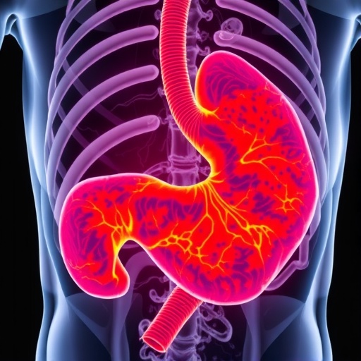
In the rapidly evolving landscape of medical diagnostics, the integration of deep learning (DL) models into pathology is heralding a new era of precision and efficiency. Gastric cancer (GC), a formidable global health challenge, demands accurate and timely diagnosis to optimize patient outcomes. Traditional histopathological examination, while effective, is inherently subjective and labor-intensive, often constrained by the variability of human interpretation. A recent systematic scoping review sheds light on how DL models are revolutionizing the analysis of gastric cancer pathology images, promising transformative impacts on clinical practice.
Histopathology, the microscopic examination of tissue to study the manifestations of disease, has long been the cornerstone of gastric cancer diagnosis. However, pathologists face restrictions including limited time, potential for oversight, and inconsistency across interpretations. DL models, particularly convolutional neural networks (CNNs), offer a computational approach that automates image analysis, enhancing reproducibility and potentially uncovering subtle features indiscernible to the human eye.
The review, adhering to rigorous PRISMA-ScR guidelines, systematically evaluated four major scientific databases: PubMed, Scopus, Web of Science, and IEEE Xplore, surveying literature up to mid-2025. Initially uncovering 520 relevant publications, the authors distilled this to 22 high-quality studies meeting stringent criteria focusing on DL applications in GC pathology image analysis.
.adsslot_jRyzrvEw5g{width:728px !important;height:90px !important;}
@media(max-width:1199px){ .adsslot_jRyzrvEw5g{width:468px !important;height:60px !important;}
}
@media(max-width:767px){ .adsslot_jRyzrvEw5g{width:320px !important;height:50px !important;}
}
ADVERTISEMENT
Among the most compelling findings is the performance of DL models in detecting gastric cancer presence within histological samples. Several models achieved accuracy rates exceeding 95%, rivaling or surpassing human expert assessments. This level of precision is particularly promising for early detection, a critical factor in improving survival rates given the aggressive nature of advanced gastric cancers.
Beyond mere detection, DL applications extend to histological classification, where distinguishing between various GC subtypes can influence treatment decisions. Deep learning systems have demonstrated proficiency in classifying complex cancer morphologies, facilitating more nuanced clinical insights. This capability points toward personalized treatment plans shaped by detailed tumor profiling instead of broad categories.
Prognosis prediction is another frontier illuminated by DL-driven image analysis. By extracting intricate patterns from pathology slides, these algorithms offer prognostic assessments that integrate morphological features with patient outcomes. This integration supports oncologists in stratifying patient risk and tailoring therapies more effectively, potentially improving survivorship.
CNNs dominate the current landscape of DL architectures applied in gastric cancer pathology. Their hierarchical feature extraction mechanisms, inspired by the organization of the visual cortex, make them particularly suited for the complex textures and structures characteristic of tissue images. These models excel at identifying local and global image features critical for accurate classification.
Despite impressive advancements, the review underscores significant challenges limiting clinical translation. Chief among these is the paucity of large, diverse datasets necessary to train robust DL models. Many studies relied on relatively small cohorts, raising concerns about overfitting and model generalizability. This bottleneck underscores the urgent need for collaborative data-sharing initiatives and the establishment of comprehensive, multicenter repositories.
External validation, a cornerstone of scientific credibility, remains underutilized in current research. Without testing models on independent datasets from varied clinical settings, their reliability across populations with differing genetic and environmental backgrounds remains uncertain. This gap must be addressed to ensure DL systems are broadly applicable and equitable.
Moreover, existing studies often fall short in covering the full spectrum of gastric cancer types and disease stages. Gastric cancer is biologically heterogeneous, with diverse histological patterns and clinical trajectories. Effective DL models must therefore accommodate this heterogeneity to be truly transformative in real-world clinical scenarios.
The review highlights an emerging consensus that future research should prioritize dataset expansion—not just in quantity but in quality, comprehensiveness, and representativeness. Integration of multi-institutional data, inclusion of rare subtypes, and incorporation of longitudinal clinical information will be key progress markers.
Clinical validation is also paramount. Prospective studies and clinical trials assessing the impact of DL-assisted pathology on diagnostic accuracy, turnaround times, and patient outcomes will determine the practical utility of these technologies. This phase of research is critical to moving beyond algorithm development to full implementation.
Ethical considerations arise alongside these technical challenges. Transparency in model decision-making, avoidance of biases, and maintaining patient privacy during data collection and processing are essential components in gaining clinician and patient trust.
Furthermore, the technological ecosystem surrounding DL in pathology must evolve to support integration into existing workflows. User-friendly interfaces, interoperability with digital pathology systems, and robust performance in diverse clinical environments will facilitate adoption.
Ultimately, the convergence of artificial intelligence and pathology holds the promise of democratizing expert diagnostic capabilities, enabling resource-limited settings to access advanced cancer detection tools. This vision aligns with global health objectives targeting early cancer diagnosis and treatment equity.
As the field progresses, interdisciplinary collaboration among computer scientists, pathologists, oncologists, and bioinformaticians will be key. Combining domain expertise with computational innovation will refine algorithms and ensure clinical relevance.
In conclusion, deep learning models are poised to revolutionize gastric cancer pathology image analysis, offering unprecedented accuracy in detection, classification, and prognosis prediction. To fully unlock this potential, future research must surmount current limitations through expanded datasets, rigorous external validations, and comprehensive clinical assessments. These strides promise to enhance patient care and reshape the future of cancer diagnostics.
Subject of Research: Application of deep learning models in gastric cancer pathology image analysis.
Article Title: Application of deep learning models in gastric cancer pathology image analysis: a systematic scoping review.
Article References:
Xia, S., Xia, Y., Liu, T. et al. Application of deep learning models in gastric cancer pathology image analysis: a systematic scoping review. BMC Cancer 25, 1257 (2025). https://doi.org/10.1186/s12885-025-14662-3
Image Credits: Scienmag.com
DOI: https://doi.org/10.1186/s12885-025-14662-3
Tags: accuracy in gastric cancer detectionadvances in histopathology techniquesautomated image analysis for gastric cancerconvolutional neural networks in pathologydeep learning models in gastric cancer diagnosisenhancing reproducibility in cancer diagnosisimproving patient outcomes with technologymachine learning applications in oncologyovercoming human bias in pathologyprecision medicine in cancer treatmentsystematic review of DL in medical diagnosticstransformative impact of AI on healthcare


