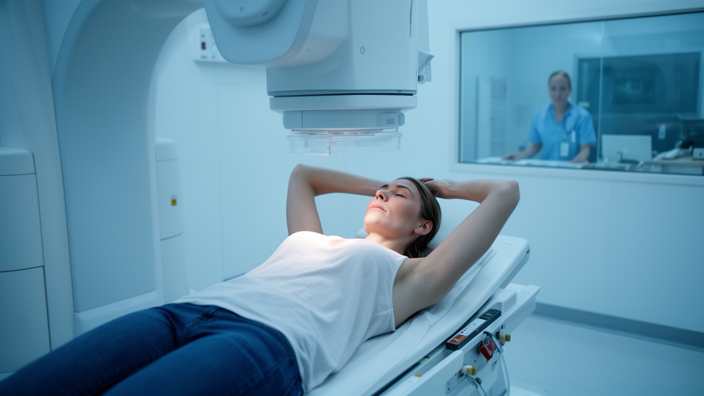
For women treated for left-sided breast cancer, one of the most persistent worries is not the tumor itself but the collateral damage that radiation can inflict on the heart and lungs lying just beneath the treatment field. A new study from Xingtai People’s Hospital in China offers fresh quantitative evidence that a simple breathing maneuver—holding a deep breath during each radiation session—can meaningfully cut the dose delivered to these vulnerable organs, and it goes a step further by probing why some patients benefit more than others.
The technique, known as deep inspiration breath-hold, or DIBH, is not new. When a patient inhales deeply and holds her breath, the lungs expand, the diaphragm drops, and the heart is physically pulled away from the chest wall and out of the path of the radiation beam. The effect is geometric rather than pharmacological: by shifting anatomy, clinicians can spare healthy tissue without weakening the treatment aimed at the breast. What has remained murky is the degree to which individual anatomy determines how much protection each patient actually gains.
In the study, published in Frontiers in Oncology, a team led by Yanli Cui and Shuai Xu enrolled 17 patients with stage I left-sided breast cancer who had undergone breast-conserving surgery between July 2023 and July 2024. Each patient underwent two separate CT simulation scans—one while breathing freely and one while performing DIBH with the help of an Active Breathing Coordinator system, which monitors airflow and locks the airway at a preset inspiratory volume. From each scan, the team generated a full intensity-modulated radiation therapy plan using identical prescription doses and optimization objectives, allowing a direct, within-patient comparison of how the same treatment would deposit dose under the two breathing conditions.
The dosimetric results were consistent and statistically significant across the board. The mean dose to the left lung fell from 12.69 Gy under free breathing to 11.61 Gy with DIBH, while the volumes of lung receiving 20 Gy (V20) and 30 Gy (V30) also dropped significantly. The heart fared even better: mean heart dose declined from 5.83 Gy to 5.02 Gy, and the volumes receiving 20, 30, and 40 Gy were all significantly reduced. The benefits extended to cardiac substructures as well. The mean dose to the left ventricle fell from 7.61 Gy to 6.40 Gy, the right ventricle from 5.53 Gy to 4.58 Gy, and the left anterior descending coronary artery—the vessel most closely associated with radiation-induced cardiac events—from 22.76 Gy to 18.65 Gy.
Those numbers matter because of what earlier epidemiology has established. A landmark 2013 analysis by Sarah Darby and colleagues in the New England Journal of Medicine found that the risk of ischemic heart disease in irradiated breast cancer patients rose roughly 7.4 percent for every additional Gray of mean heart dose. Against that benchmark, a reduction of nearly 0.8 Gy in mean cardiac dose, and more than 4 Gy to the LAD, represents a potentially meaningful decrement in lifetime cardiovascular risk—though the authors of the new study are careful to note that dosimetric gains cannot be directly translated into clinical outcomes without long-term follow-up.
The more novel contribution of the work lies in its attempt to explain the variability. The researchers measured a battery of anatomical parameters on each patient’s scans: the maximum cardiac margin distance (MCMD), a measure of how close the heart comes to the chest wall near the treatment field; the three-dimensional displacement of the heart’s geometric center between the two breathing states; the total volume of cardiac displacement; the heart-to-breast distance; and the change in lung volume induced by deep inspiration. They then correlated these measurements with the dosimetric changes observed between plans.
Several relationships emerged. Under DIBH, the MCMD was positively correlated with mean heart dose, meaning patients whose hearts sat closer to the chest wall received higher doses even with the breath-hold technique. More tellingly, the change in MCMD between free breathing and DIBH was negatively correlated with heart dose parameters—patients whose hearts moved farther from the chest wall during deep inspiration tended to gain more cardiac sparing. The change in MCMD correlated negatively with mean heart dose (r = -0.516), heart V20 (r = -0.538), heart V30 (r = -0.578), heart V40 (r = -0.575), and the mean doses to both the left and right ventricles.
The heart, it turns out, does not simply retreat in one direction. The team found that during DIBH the heart shifted predominantly along the anterior-posterior axis, with displacement ranging from 2.5 to 27.6 millimeters and averaging about 9 millimeters. Displacement along that axis was itself negatively correlated with mean heart dose. The total cardiac displacement volume ranged from about 62 to 191 milliliters, and patients with larger displacement volumes showed greater reductions in mean heart dose (r = 0.689). Meanwhile, the average increase in lung volume between the two breathing states was a substantial 1,314 milliliters, and that expansion correlated with posterior cardiac displacement, with changes in heart-to-breast distance, and—negatively—with the mean dose to the LAD under DIBH.
Taken together, these findings sketch a mechanical picture of why DIBH works and why it works unevenly. Deep inspiration inflates the lungs, and the inflated lungs push the heart backward and downward, away from the tangential radiation beams that treat the breast. Patients whose anatomy permits a larger excursion—more compliant lungs, more mobile hearts, more room in the thorax—reap larger dose reductions. Those whose hearts remain close to the chest wall even at full inspiration gain less. In principle, this suggests that a quick anatomical assessment at simulation could identify which patients stand to benefit most, though the authors explicitly caution that their exploratory correlations, drawn from a small single-center cohort without correction for multiple comparisons, are hypothesis-generating rather than practice-changing.
The study’s limitations are candidly acknowledged. Seventeen patients is a small sample; the analysis was retrospective; no long-term clinical outcomes such as cardiac events or pneumonitis were assessed; and the anatomical parameters have not been validated in an independent cohort. Still, the work adds a useful layer to a growing literature that has moved beyond asking whether DIBH is effective—it clearly is—to asking which patients gain the most and why. If the correlations hold up in larger, multicenter studies, simple geometric measurements taken during routine CT simulation could become a practical tool for personalizing respiratory management in breast radiotherapy, ensuring that the patients who need cardiac protection most are the ones who get it.
Subject of Research: Dosimetric effects of deep inspiration breath-hold in postoperative radiotherapy for left-sided breast cancer
Article Title: Dosimetric benefits of deep inspiration breath-hold in postoperative radiotherapy for left-sided breast cancer after breast-conserving surgery: an anatomical parameter-based analysis
Article References: Cui, Y., Xu, S., Wang, X., Huo, L., Wen, J., Hao, L., Wang, X., Zhang, Q., & Duan, H. (2026). Dosimetric benefits of deep inspiration breath-hold in postoperative radiotherapy for left-sided breast cancer after breast-conserving surgery: an anatomical parameter-based analysis. Frontiers in Oncology, 16, Article 1917416. https://doi.org/10.3389/fonc.2026.1917416
Image Credits: AI Generated
DOI: 10.3389/fonc.2026.1917416
Keywords: deep inspiration breath-hold, left-sided breast cancer, radiotherapy, cardiac dose, lung dose, dosimetry, IMRT, breast-conserving surgery, left anterior descending coronary artery, cardiac displacement, organ at risk sparing, radiation oncology
News Source: Nathaniel Bowman. (October 19, 2026). Deep Breath During Radiotherapy Shields Heart in Left-Sided Breast Cancer. Scienmag.