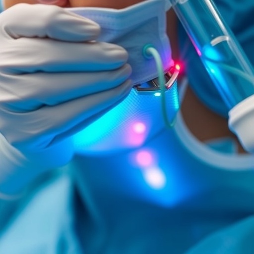
A single session of direct-current microcurrent stimulation was associated with an immediate reduction in the measured depth of postsurgical scars, according to a preliminary study using high-resolution three-dimensional ultrasound. Researchers reported that scar depth decreased by an average of 0.447 millimeters—equivalent to a mean reduction of 23.8%—after treatment. The findings, published in the Journal of Bodywork & Movement Therapies, are attracting attention because they use objective imaging to assess a structural change in scar tissue rather than relying only on photographs, visual inspection or patient impressions.
The prospective observational study involved 17 people with surgically induced scars. Each participant underwent ultrasound imaging immediately before and after one treatment session. Investigators used a 17-megahertz three-dimensional volumetric ultrasound probe to measure scar depth at the same anatomical locations at both time points. The technology produces high-resolution images of tissue architecture and allows researchers to estimate the dimensions of superficial structures, including areas of thickened or fibrotic scar tissue.
Treatment was delivered using two Dolphin Neurostim devices designed to provide direct-current microcurrent stimulation. Researchers applied the stimulation bilaterally along each scar, placing treatment points at intervals of approximately half an inch. Each point was treated for about 30 seconds, with opposing electrical polarities used on either side of the scar. A complete session lasted approximately 15 to 20 minutes. Unlike conventional electrical stimulation used to activate muscles, microcurrent protocols typically operate at low intensities intended to influence local tissue without producing visible muscle contraction.
The primary finding was a statistically significant reduction in ultrasound-measured scar depth immediately after treatment. The reported 95% confidence interval for the average change ranged from 0.264 to 0.630 millimeters, while the statistical significance level was p<0.001. In practical terms, the result suggests that the measured tissue profile became shallower during the short interval between the pre-treatment and post-treatment scans. However, the study design cannot determine whether the reduction reflects lasting remodeling of collagen-rich tissue, temporary changes in hydration or tissue compression, altered mechanical tension, or a combination of these factors.
Scar tissue, also known as cicatrix, forms when the body repairs damaged tissue through the deposition and reorganization of collagen. Mature scars can become thickened, rigid or tethered to deeper layers, potentially restricting movement between the skin, fascia and underlying structures. These adhesions may contribute to a sensation of tightness or mechanical discomfort. The researchers propose that microcurrent stimulation could influence local tissue behavior and scar morphology, but the biological mechanism remains uncertain and was not directly tested in the study.
The authors describe the work as the first report, to their knowledge, of direct-current microcurrent therapy producing a measurable reduction in the physical size of scar tissue. The use of three-dimensional ultrasound is central to that claim. High-frequency ultrasound can distinguish changes in tissue boundaries and thickness without invasive sampling, making it possible to examine treatment responses repeatedly. Still, imaging measurements can be affected by probe pressure, probe angle, the exact location of the scan and the way tissue boundaries are identified. Standardized scanning and blinded assessment will therefore be important in future research.
Participants also spontaneously reported improvements in pain and range of motion after treatment. Those experiences are potentially relevant because scar tethering can interfere with normal movement and may create mechanical irritation during rehabilitation. However, pain and mobility were not formally measured using validated scales or objective motion testing. The reports consequently cannot establish that the treatment improved function, and they may have been influenced by expectation, immediate tissue manipulation or the natural variability of symptoms.
The study’s preliminary nature places important limits on the interpretation of its viral headline result. It had no untreated control group, sham-treatment group or randomization, and measurements were taken only immediately after one intervention. Without a comparison group, researchers cannot determine how much of the observed change was caused by microcurrent stimulation itself or by factors such as positioning, scanning pressure or ordinary measurement variability. The study also does not show whether the scar remained shallower hours, days or weeks later, or whether repeated treatments would produce cumulative benefits.
The researchers say larger controlled trials with longer follow-up are needed to test the durability and clinical importance of the finding. Future studies could compare different current intensities, treatment frequencies and polarity arrangements while using blinded ultrasound analysis and standardized assessments of pain, range of motion, scar pliability and patient function. Additional imaging or tissue studies might also help determine whether any immediate change represents true collagen remodeling or a reversible alteration in tissue mechanics. For now, the study offers an intriguing imaging-based signal rather than definitive evidence that microcurrent therapy permanently removes or remodels surgical scars.
Subject of Research: People
Article Title: Effects of DC Microcurrent Stimulation on Scar Remodeling and Tissue Recovery
News Publication Date: August 12, 2026
Web References: https://doi.org/10.1016/j.jbmt.2026.07.039
References: Gokal R, Armstrong K, Todorsky W, Durant J. Effects of DC Microcurrent Stimulation on Scar Remodeling and Tissue Recovery. Journal of Bodywork & Movement Therapies. DOI: 10.1016/j.jbmt.2026.07.039.
Keywords
DC microcurrent stimulation, scar remodeling, postsurgical scars, three-dimensional ultrasound, tissue recovery, fibrosis, cicatrix, biomedical engineering, rehabilitation, pain research
Tags: clinical study on microcurrent therapydirect-current stimulation for tissue repaireffects of neurostimulation devices on scar depthhigh-resolution 3D ultrasound in wound analysisinnovative approaches to scar managementmicrocurrent stimulation for scar healingminimally invasive scar reduction methodsnon-invasive scar treatment techniquesobjective measurement of scar tissue changespostsurgical scar reductionstructural changes in scars post-treatmentultrasound imaging in scar assessment