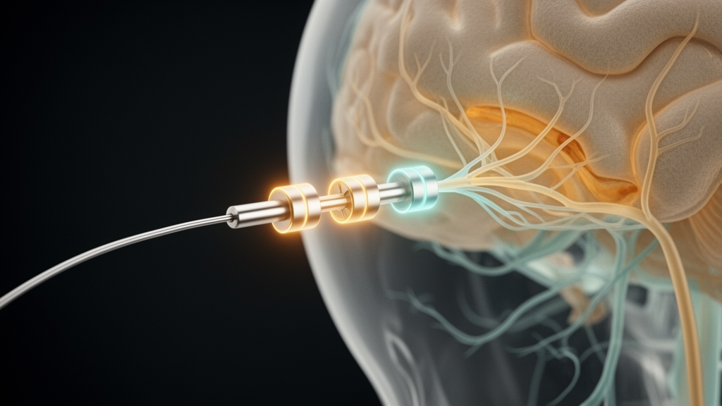
A retrospective study published in Frontiers in Human Neuroscience has identified modest but systematic spatial differences between the optimal stimulation sites identified during surgery and those effective in the postoperative phase for patients with Essential Tremor. The research, led by Vittoria Bucciarelli and colleagues, investigated whether the ‘Probabilistic Sweet Spots’ (PSSs) mapped from intraoperative data remain spatially stable over time. The findings suggest that while there is a considerable overlap between the two phases, the optimal stimulation regions are not identical, with a consistent anterior-posterior displacement observed in the left hemisphere.
Deep Brain Stimulation is an established therapy for movement disorders, including Essential Tremor and Parkinson’s Disease. In clinical practice, stimulation testing is performed at multiple time points to optimize electrode placement and parameters. Intraoperative testing is widely used to guide implantation, allowing for the identification of stimulation locations associated with optimal clinical benefit. If these changes occur, stimulation settings inferred from intraoperative data may not remain valid in the postoperative phase, potentially limiting their usefulness for early programming. This issue is particularly relevant as the trend shifts toward DBS procedures performed under general anesthesia, where awake intraoperative stimulation testing is not feasible.
The study analyzed data from 14 patients with Essential Tremor who underwent bilateral DBS implantation at the Department of Neurosurgery of Clermont-Ferrand University Hospital in France between 2008 and 2018. The electrodes targeted the ventrointermediate nucleus of the thalamus (Vim) for tremor alleviation. The researchers retrospectively analyzed 324 intraoperative and 566 postoperative stimulation tests. Postoperative testing was conducted approximately three months after surgery to minimize the influence of lesioning effects. The analysis was conducted on two levels: a cohort level using group analysis and a patient-specific basis, allowing for both statistical testing and the preservation of individual variability.
To determine the optimal stimulation sites, the team employed electric field simulations for all tested stimulation settings. Using the ELMA1 software, they generated patient-specific brain conductivity models and simulated the electric field in the brain tissue. Volumes of Tissue Activated (VTAs) were delineated by thresholding the electric field at 0.2 V/mm. Probabilistic Sweet Spots were then computed by applying voxel-wise statistical testing to these VTAs. At the group level, a Bayesian t-test was used to identify voxels where the tremor improvement score exceeded specific thresholds, such as 12.5%, 25%, 50%, and 75%. At the individual patient level, a descriptive voxel-wise approach was adopted, using a MedianMap to represent the median improvement scores of all VTAs stimulating each voxel.
At the group level, the study observed a partial spatial concordance between intraoperative and postoperative PSSs. The highest agreement between phases was observed at the 12.5% improvement threshold, with a Dice coefficient of 0.75 in the left hemisphere and 0.58 in the right hemisphere. However, this overlap decreased monotonically as the improvement threshold increased. At the 50% threshold, the Dice coefficient dropped to 0.18 in the left hemisphere and 0.43 in the right hemisphere.
At the individual patient level, the analysis revealed a consistent anterior-posterior displacement in the left hemisphere. Intraoperative PSSs were located more posteriorly than postoperative PSSs, with a mean displacement of approximately 1.5 mm. This shift was statistically significant along the anterior-posterior orientation. In contrast, no significant differences in centroid positions were detected in the right hemisphere along any of the three orientations. The intraoperative PSS volumes were also significantly smaller than postoperative volumes overall, with a mean intraoperative volume of 296 mm³ compared to a mean postoperative volume of 477 mm³. This difference was particularly pronounced in the right hemisphere, where postoperative volumes were significantly larger than intraoperative volumes.
The researchers investigated whether these shifts were correlated with displacements between the intraoperative and postoperative electrode trajectories. The analysis showed that shifts between intraoperative and postoperative PSSs did not show a significant correlation with corresponding trajectory shifts. However, a positive trend was observed for shifts along the right-left and anterior-posterior orientations. The authors suggest that the observed displacements may reflect both genuine spatial changes and methodological differences between the two assessments. Intraoperative testing used a 0.5 mm diameter microelectrode with current-controlled stimulation, while postoperative testing used a 1.27 mm diameter chronic electrode with voltage-controlled stimulation. These differences in electrode geometry and sampling strategies may have contributed to the observed PSS displacements.
The study highlights the importance of considering temporal evolution in Probabilistic Sweet Spot analyses. Previous studies typically relied on a single chronic time point, most often one year after implantation, and did not address the temporal evolution of PSSs. By incorporating previously unpublished postoperative stimulation data, this work enables a direct temporal comparison of PSSs across surgical phases. The findings indicate that modest spatial differences arise between intraoperative and postoperative PSSs, likely attributable to brain shift, differences in electrode characteristics, and postoperative tissue changes. The magnitude of these displacements remains in the order of a couple of millimeters, which is within the limits of surgical precision.
The authors note that the high overlap between intraoperative and postoperative PSSs suggests that probabilistic spatial mapping may complement conventional threshold-based assessments. This approach provides anatomically informed guidance for postoperative programming, potentially reducing the need for extensive early patient screening. However, the study has several limitations. The relatively small patient cohort of 14 individuals may limit statistical power and generalizability. Additionally, the imbalance between the number of postoperative and intraoperative tests, with postoperative testing being more extensive, may have influenced the results. The different number of stimulation tests, together with differences in electrode geometry and sampling density, may have contributed to the observed PSS displacements.
Furthermore, although postoperative CT was used to verify lead location following implantation, no additional imaging was performed immediately before the three-month postoperative assessment. Consequently, small changes in lead position between implantation and postoperative testing cannot be excluded. Brain shift may also have contributed to discrepancies between planned and achieved lead positions. These factors represent additional sources of uncertainty in the spatial comparison of intraoperative and postoperative PSSs. The selection of improvement thresholds for PSS computation also remains a source of variability in the workflow, introducing methodological uncertainty that warrants further standardization in future studies.
Despite these limitations, the study provides valuable insights into the spatial stability of optimal stimulation sites in DBS for Essential Tremor. The findings suggest that intraoperative PSSs may inform early postoperative DBS programming, supporting and streamlining early postoperative screening. However, whether PSS locations remain stable over longer follow-up periods remains an open question. Longer-term evaluations at one or two years could provide additional insight into the mechanisms of DBS, the evolution of stimulation responses over time, and disease progression.
The research underscores the complexity of translating intraoperative findings to the chronic stimulation context. While the observed shifts are modest, they are systematic and may have clinical implications for the optimization of DBS parameters. The use of electric field simulations and probabilistic mapping offers a robust framework for characterizing the spatial stability of therapeutic stimulation regions. By integrating voxel-wise efficacy information, this approach enables a more detailed understanding of how optimal stimulation sites evolve over time.
Subject of Research: Spatial stability of Probabilistic Sweet Spots in Deep Brain Stimulation for Essential Tremor
Article Title: Assessing systematic spatial differences between intraoperative and postoperative Probabilistic Sweet Spots in Deep Brain Stimulation for Essential Tremor
Article References: Bucciarelli, V., Vogel, D., Coste, J., Lemaire, J.-J., Derost, P., Debilly, B., Guzman, R., & Hemm, S. (2026). Assessing systematic spatial differences between intraoperative and postoperative Probabilistic Sweet Spots in Deep Brain Stimulation for Essential Tremor. Frontiers in Human Neuroscience, 20, Article 2005438. https://doi.org/10.3389/fnhum.2026.2005438
Image Credits: AI Generated
DOI: 10.3389/fnhum.2026.2005438
Keywords: Deep Brain Stimulation, Essential Tremor, Probabilistic Sweet Spots, Neuroscience, Medical Imaging, Clinical Neurology, Stimulation Therapy, Retrospective Study, Assessing, systematic, spatial, differences
News Source: Ophelia Keating. (November 10, 2026). DBS Sweet Spots Shift Slightly After Essential Tremor Surgery. Scienmag.