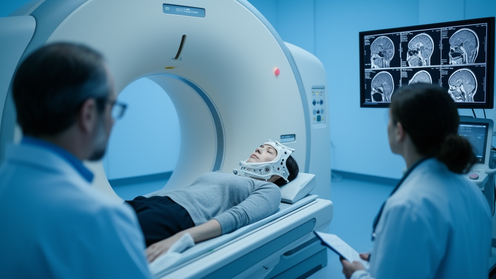
Every day, millions of patients undergoing radiotherapy pass through the beam of a cone-beam computed tomography scanner, an imaging device mounted on the treatment machine itself. Its purpose is simple: confirm that the tumour is precisely aligned with the radiation field before each fraction is delivered. Yet those daily scans contain far more information than positioning checks alone. A team of researchers at the Lithuanian University of Health Sciences in Kaunas has now shown that, with the right computational framework, routine cone-beam CT images can be transformed into a longitudinal record of how a tumour’s internal texture changes week by week as radiation does its work. Their study, published in BMC Medical Imaging, offers a methodological blueprint for turning an underused by-product of radiotherapy into a potential window on treatment response.
The research focuses on head and neck squamous cell carcinoma, one of the most heterogeneous malignancies in clinical oncology. Patients with seemingly similar tumours can respond to identical radiation prescriptions in dramatically different ways, and clinicians currently have few tools for detecting those differences while treatment is still underway. Conventional assessment typically relies on imaging performed before and after the full course of therapy, a comparison that captures the outcome but misses the trajectory. The Kaunas group set out to characterise what happens in between, using the cone-beam CT scans that are already acquired as part of standard patient positioning.
The central technical challenge is one of consistency. Cone-beam CT images are acquired daily with the patient in the treatment position, but the tumour does not sit in exactly the same place from one fraction to the next. Patients lose weight, anatomy shifts, and the tumour itself shrinks or deforms. Before any meaningful comparison of tumour appearance across time can be made, the volumes of interest must be brought into spatial correspondence. The researchers addressed this by defining tumour volumes on the planning CT and propagating them to each daily cone-beam image using rigid point-cloud registration, a technique that aligns sets of anatomical points so that the same region of tissue is analysed at every fraction. This registration-consistent approach ensures that changes in measured texture reflect biology rather than shifting geometry.
Within those longitudinally aligned volumes, the team extracted radiomic features, quantitative descriptors that summarise the statistical texture of an image. Radiomics rests on the idea that the spatial arrangement of pixel intensities encodes biological properties such as cell density, necrosis, and tissue heterogeneity. Three features anchored the analysis: entropy, a measure of randomness or disorder in the image; strength, which characterises how strongly neighbouring voxels differ in intensity; and coarseness, which captures the size of the texture elements, with coarser textures corresponding to larger, more uniform regions. Each of these descriptors was computed at every treatment fraction, producing a time series for every patient.
To model the resulting trajectories, the researchers turned to linear mixed-effects models, a statistical framework well suited to repeated-measures data. Mixed-effects models can separate the overall population trend from individual patient deviations, and they accommodate unbalanced data, an important consideration when not every patient has the same number of usable scans. The models incorporated treatment fraction as a temporal variable, quadratic terms to capture nonlinear dynamics, and tumour differentiation group, graded from G1 to G3, as a potential modifier of the temporal patterns. This allowed the team to ask not only whether features change over time, but whether the shape of that change differs according to tumour biology.
The results revealed a striking divergence in robustness among the three features. Entropy, the measure of textural disorder, displayed significant nonlinear temporal behaviour with a p-value below 0.001, characterised by a mid-treatment peak. In other words, tumour texture appeared to become most disordered partway through the course of radiotherapy before evolving further toward the end. But entropy also showed substantial variability between patients, meaning that individual trajectories could deviate considerably from the population average. That variability complicates any attempt to read entropy values as a straightforward indicator of response in a single patient.
Strength, the second feature, exhibited only modest nonlinear variation over the course of treatment, and the analysis found no significant effects associated with tumour differentiation group. Its trajectory, while measurable, offered neither the dynamic range of entropy nor the stability of coarseness. The third feature, coarseness, emerged as the standout. It demonstrated the most consistent longitudinal behaviour of the three, with significant group-dependent temporal patterns at a significance level below 0.01 and well-defined turning points that differed across the G1 to G3 differentiation groups. Crucially, coarseness trajectories were less affected by high-frequency variation than entropy or strength, suggesting that the feature is less susceptible to the noise and artefacts that plague cone-beam CT imaging.
That robustness matters because cone-beam CT is not a diagnostic-grade scanner. Its images suffer from scatter, limited contrast, and dose constraints, all of which can corrupt fine textural measurements. A radiomic feature intended for clinical use must survive those imperfections. The finding that coarseness, a descriptor of large-scale texture organisation, remains stable and interpretable across fractions while finer-grained measures wobble is consistent with the intuition that coarse texture is less sensitive to high-frequency imaging noise. It also aligns with the biology: as radiation kills tumour cells, tissue architecture changes at scales that coarseness is well positioned to detect.
The authors are careful about what these findings do and do not support. The study was retrospective, analysing thirty patients treated with definitive radiotherapy, and it did not link the measured texture trajectories to clinical endpoints such as survival or locoregional control. The team explicitly states that prospective validation with clinical outcomes is required before any biomarker claims can be made. What the work delivers instead is a methodological foundation: a registration-consistent framework for aligning daily images, a demonstration that longitudinal radiomic trajectories can be modelled rigorously with mixed-effects statistics, and an evidence-based ranking of which features deserve priority in future validation studies.
If that validation succeeds, the implications for radiotherapy could be substantial. Adaptive radiotherapy, the practice of modifying treatment plans mid-course in response to observed changes, currently depends largely on anatomical measurements such as tumour volume. Texture-based descriptors could add a functional dimension, potentially flagging patients whose tumours are responding unusually quickly or slowly while there is still time to intervene. Because the required images are already acquired daily at essentially every modern radiotherapy facility, the marginal cost of such monitoring would be minimal. The Kaunas framework suggests that the data needed to personalise head and neck cancer treatment may already be sitting in the scanner, waiting to be read.
Subject of Research: Longitudinal CBCT radiomics for monitoring treatment-induced tumour texture changes in head and neck radiotherapy
Article Title: A registration-consistent longitudinal CBCT framework for quantifying treatment-induced tumour texture changes in head and neck radiotherapy
Article References: Karpaviciene, G., Meilutyte-Lukauskiene, D., Cerapaite-Trusinskiene, R., Paukstaitiene, R., Speckauskiene, V., & Petrolis, R. (2026). A registration-consistent longitudinal CBCT framework for quantifying treatment-induced tumour texture changes in head and neck radiotherapy. BMC Medical Imaging. https://doi.org/10.1186/s12880-026-02909-9
Image Credits: AI Generated
DOI: 10.1186/s12880-026-02909-9
Keywords: cone-beam CT, head and neck cancer, radiotherapy, radiomics, delta radiomics, tumour heterogeneity, image registration, mixed-effects modelling, treatment response, adaptive radiotherapy, imaging biomarkers, medical imaging
News Source: Nathaniel Bowman. (October 9, 2026). Daily CT Scans Reveal Hidden Tumour Texture Shifts During Head and Neck Radiotherapy. Scienmag.