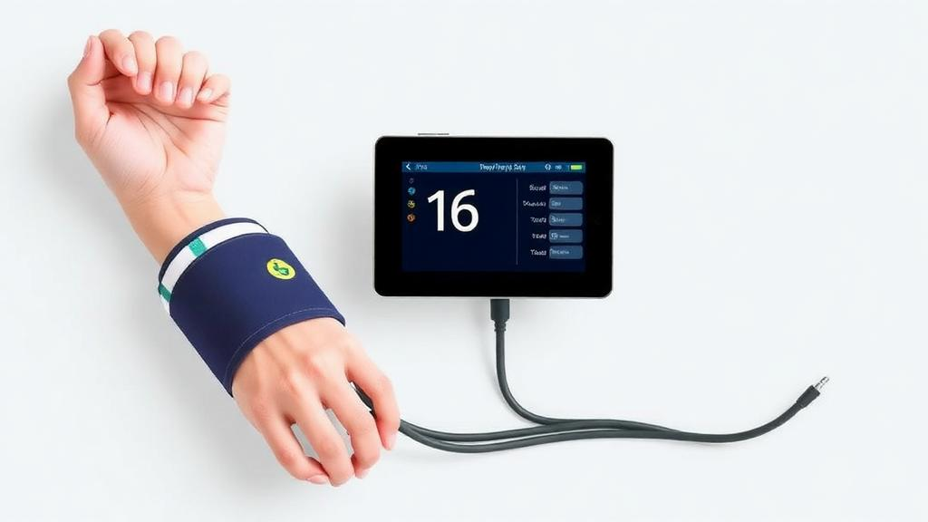
Peripheral artery disease, the silent narrowing of the arteries that carry blood to the arms and legs, affects hundreds of millions of people worldwide and remains one of the most underdiagnosed vascular conditions in medicine. Now, a team of biomedical engineers and clinicians in Taiwan has built and tested a device that could change how the disease is screened: a cuffless platform that clips onto all four limbs at once, synchronously records the heart’s electrical activity together with optical pulse waveforms from each limb, and analyzes the timing differences to flag blocked or stiffened arteries. The work, published in the Journal of Medical and Biological Engineering by Ming-Jui Wu of Tajen University and Kaohsiung Veterans General Hospital Tainan Branch, together with I-Hua Huang and Yi-Chun Du of National Cheng Kung University, targets a diagnostic blind spot that has frustrated vascular specialists for decades.
The standard screening tool for peripheral artery disease is the ankle–brachial index, a simple ratio obtained by measuring blood pressure at the ankle with an inflatable cuff and dividing it by the pressure at the arm. A low ratio signals that blood flow to the lower limbs is compromised. The test is cheap and widely used, but it has a critical weakness: in patients whose ankle arteries have become stiff and calcified, the vessels resist compression by the cuff, the measured ankle pressure reads artificially high, and the index appears deceptively normal. This problem is most severe in people with chronic kidney disease, where medial arterial calcification is common and the sensitivity of the ankle–brachial index falls to roughly 43 percent. In other words, more than half of diseased patients in this high-risk group can walk out of a screening clinic with a clean bill of health.
The Taiwanese team’s answer is to abandon the cuff altogether. Their platform measures pulse transit time, the small delay between the electrical trigger of a heartbeat, captured by an electrocardiographic electrode, and the arrival of the resulting pressure pulse at a peripheral site, detected optically with photoplethysmography. Because arterial stiffness and obstruction slow and distort the traveling pulse, these transit delays carry information about the state of the vasculature between the heart and the measurement point. Crucially, the device records all four limbs simultaneously against a single cardiac reference, something that existing systems typically achieve only by chaining together separate sensors that cannot be trusted to share a common clock.
The engineering solution is elegantly compact. Each limb clip carries both a dry electrocardiographic electrode and a transmission-mode photoplethysmographic sensor on a single contact point, so attaching the device requires no gel, no cuff inflation, and no separate electrode placement. One electrocardiographic channel and four photoplethysmographic channels are digitized together at 500 hertz, ensuring that every waveform is sampled on the same time base. Before any feature is extracted, the system applies a quality gate: an optical channel is accepted only when its pulses occur at the same rate as the electrocardiogram, filtering out motion artifacts and poor contact. Features are then computed only from heartbeats that can be resolved cleanly at all four limbs at once, a discipline that trades raw data volume for reliability.
Bench testing confirmed that the hardware meets the precision demands of transit-time measurement. The platform achieved 35.9 decibels of mains interference rejection, important because power-line noise at 50 or 60 hertz can swamp the microvolt-level electrocardiographic signal. The photoplethysmographic passband gain reached 352-fold, amplifying the faint optical flicker of blood volume changes in the fingertip or toe, and reproducibility between boards was within plus or minus 2 percent, meaning different units of the device would produce comparable measurements. In the human recordings, the median pulse transit time to the feet was 83 milliseconds longer than to the hands, a physiological difference that reflects the greater distance the pressure wave must travel and that confirms the system can resolve meaningful spatial gradients across the body.
The clinical pilot involved twenty-three participants, six of whom had both peripheral artery disease and chronic kidney disease, the very population in which conventional screening fails most badly. The results were striking. Pulse transit time measured at the toe correlated strongly with the reference ankle–brachial index, with a correlation coefficient of negative 0.88, meaning that as the index fell, indicating worse disease, the optical transit delay grew longer. More importantly, the platform statistically separated the patients with both peripheral artery disease and chronic kidney disease from the control participants with a p-value below 0.001, a discrimination that the ankle–brachial index itself could not achieve in this cohort. Because the optical method detects the pulse as it passes through tissue without compressing the artery, calcified vessels cannot fool it the way they fool a cuff.
The practical advantages extend beyond diagnostic accuracy. Setting up the device took less than a minute and required no cuff inflation, which makes it plausible for point-of-care screening in clinics, dialysis units, or even community health settings where chronic kidney disease patients are already being monitored. Dry electrodes eliminate the skin preparation and conductive gel that slow down conventional electrocardiographic setups, and the single-contact clip design means a trained technician could apply the sensors quickly and symmetrically to all four limbs. For a disease that is often asymptomatic until it reaches an advanced stage, a screening tool that is fast, comfortable, and reliable in high-risk populations could shift the detection curve substantially.
The study is candid about its main limitation: signal yield. The optical channels performed very differently at different sites. Segments recorded at the hands passed the quality gate 91 percent of the time, but at the feet the pass rate dropped to 54 percent, a statistically significant difference, and when the analysis demanded clean signals at all four limbs simultaneously, only 41 percent of segments qualified. The toes, with their smaller vessels and thinner perfusion, produce weaker and noisier photoplethysmographic signals, particularly in older patients and those with vascular disease, precisely the people the device is meant to help. The authors identify raising the optical sensitivity at the feet as the principal engineering route to a higher four-limb yield, and the small sample size means the clinical findings will need validation in larger, prospectively designed cohorts before the platform could approach regulatory clearance or routine clinical use.
Even so, the work lands at a moment of intense interest in cuffless vascular assessment. Photoplethysmography has exploded beyond the clinic into smartwatches and fitness bands, and researchers worldwide are mining the pulse waveform for information about arterial stiffness, blood pressure, and vascular age. Most of that effort relies on single-site or two-site measurements, however, and the four-limb synchronous architecture demonstrated here offers something those approaches cannot: a direct, simultaneous comparison of pulse timing between the arms and legs against one cardiac reference, which is exactly the physiological contrast that defines peripheral artery disease. Related efforts using multi-site photoplethysmography and machine learning for lower extremity artery detection are appearing rapidly in the literature, suggesting the field is converging on this measurement strategy from several directions at once.
If the yield problems at the feet can be solved, the implications for patients with chronic kidney disease could be profound. This population faces a doubly elevated risk: kidney disease accelerates both atherosclerosis and medial calcification, so they are more likely to develop peripheral artery disease and less likely to have it detected by the standard test. A screening method that sidesteps the calcification trap, requires no cuff, and delivers a result in under a minute could be deployed in dialysis centers where these patients already spend hours each week. The Taiwanese platform is an early prototype rather than a finished product, but it demonstrates that the physics of the pulse, read optically and timed electrically, can succeed where the blood pressure cuff fails, and it sketches a credible path toward screening that reaches the patients who need it most.
Subject of Research: A four-limb synchronous ECG and photoplethysmography platform for cuffless screening of peripheral artery disease
Article Title: A Four-Limb Synchronous ECG and PPG Acquisition and Analysis Platform for Cuffless Peripheral Artery Disease Screening
Article References: A Four-Limb Synchronous ECG and PPG Acquisition and Analysis Platform for Cuffless Peripheral Artery Disease Screening. (n.d.). https://doi.org/10.1007/s40846-026-01058-2
Image Credits: AI Generated
DOI: 10.1007/s40846-026-01058-2
Keywords: peripheral artery disease, photoplethysmography, electrocardiography, pulse transit time, ankle–brachial index, chronic kidney disease, arterial calcification, cuffless screening, biomedical engineering, point-of-care diagnostics, vascular stiffness, signal quality
News Source: Ophelia Keating. (October 14, 2026). Cuffless Four-Limb Sensor Platform Screens for Peripheral Artery Disease Without a Blood Pressure Cuff. Scienmag.