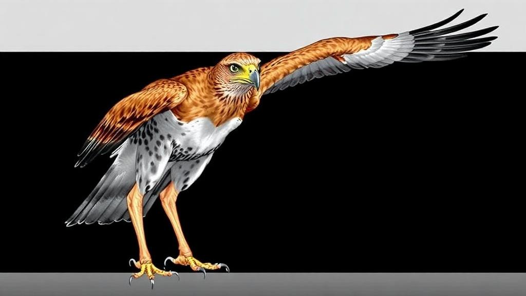
The long-legged buzzard is one of the most striking raptors of the dry steppes and mountain margins of western Asia, a bird whose body stretches up to 65 centimetres and whose wings span nearly a metre and a half. Yet for all its aerial mastery, this apex predator is remarkably vulnerable to one of the most mundane hazards of veterinary medicine: the fracture. Trauma is the leading reason birds of prey arrive at wildlife rehabilitation clinics, and the humerus, the great bone of the wing, is among the most frequently broken. Now, a team of researchers in Iran has produced the first detailed imaging reference for the humerus of Buteo rufinus, comparing digital radiography with computed tomography in ten live birds, and the results make a compelling case that the humble X-ray, for all its usefulness, sees only part of the story.
The study, conducted at a protected wildlife rehabilitation centre in Kerman province, examined ten mature long-legged buzzards weighing between 650 and 750 grams. Because the species is monomorphic, meaning males and females cannot be distinguished by external appearance, and because the birds were imaged outside the breeding season with no active gonads visible on radiographs, the sex of each animal remained undetermined. An avian specialist confirmed all ten were mature based on physical characteristics, though precise ages could not be established. Each bird received a physical examination to rule out health complications that might skew the results, and body weights were measured individually on a digital balance so that anaesthetic doses could be calculated with precision.
Imaging required careful sedation with a combination of ketamine at 20 milligrams per kilogram and diazepam at 1 milligram per kilogram, administered intramuscularly to minimise stress and movement. The researchers then captured two standard orthogonal radiographic views of each wing, a medio-lateral projection and a ventro-dorsal projection from the coelomic cavity, using a direct digital radiography system set at 60 kilovoltage peak and 2.5 milliamperage-seconds with a source-to-detector distance of 90 centimetres. The birds were placed in dorsal recumbency with their wings extended, and all images were archived in a picture-archiving and communication system for later analysis.
Computed tomography followed on a 16-slice scanner, with the animals again positioned in dorsal recumbency and their wings and legs secured with microporous surgical tape. The CT protocol used 120 kilovoltage peak and 150 milliamperage-seconds, a rotation time of one second, a slice thickness of just 1 millimetre and a pitch of 1. After scanning, the images were reconstructed into bone planes on a specialised workstation, allowing the team to interrogate the humerus in cross-section in a way no plain radiograph can match. Eight parameters were then measured by a single observer across both modalities: the cranio-caudal diameter of the bone, the diameter of the medullary cavity, cortical thickness, total proximal-to-distal length, soft tissue thickness at three regions along the bone, and bone density expressed both as pixel values on radiographs and Hounsfield units on CT.
The headline finding is a stark demonstration of how much conventional radiography can mislead. On plain films, the medullary cavity of the humerus appeared to average 7.32 millimetres in diameter, while CT revealed the true figure to be just 5.67 millimetres. Cortical thickness told the mirror-image story: radiography suggested a cortex of only about 1.06 millimetres, whereas CT measured it at 1.86 millimetres, nearly 75 percent thicker. The explanation lies in the fundamental physics of projection radiography. Because an X-ray compresses a three-dimensional structure into a two-dimensional image, overlapping anatomical structures blur the boundary between dense cortical bone and the air-filled cavity within, systematically overestimating the medullary space and underestimating the cortex. For a surgeon planning fracture repair, that difference is not academic; it determines implant selection and the feasibility of intramedullary pinning.
Not every measurement diverged. The cranio-caudal diameter of the bone itself, around 9.4 millimetres by CT and 9.46 millimetres by radiography, showed only a negligible difference between the two techniques, and this was the one parameter where the comparison did not reach statistical significance. But for medullary cavity diameter, cortical thickness, total bone length, and soft tissue thickness at the proximal, middle and distal regions, the differences between modalities were all statistically significant, with CT consistently delivering the more precise values. Total humeral length, for instance, measured 140.68 millimetres on radiographs but 132.84 millimetres on CT, a discrepancy the authors attribute to the magnification and superimposition inherent in two-dimensional projection imaging.
Perhaps the most biologically intriguing numbers came from the density measurements. On CT, the cortex registered approximately 822 Hounsfield units, the medullary cavity around negative 805 Hounsfield units, and the surrounding soft tissue about 66 Hounsfield units, with no meaningful difference between the right and left wings for any parameter. The medullary value is the giveaway: a strongly negative Hounsfield reading indicates air, confirming the extensive pneumatisation that defines the avian humerus. In birds, the humerus is not filled with marrow in the mammalian sense but with air-filled cavities connected to the respiratory system, an adaptation that lightens the skeleton for flight and assists in respiration and air humidification. The researchers note that a Hounsfield value of 800 plus or minus 50 in the cortex reflects the degree of this pneumatisation, and the symmetry between wings suggests a tightly regulated developmental programme.
That pneumatisation is a double-edged sword. By hollowing the bone, evolution has made pneumatic bones more prone to shattering and fragmenting on impact, and the distal extremities of the humerus lack substantial soft tissue protection, leaving them vulnerable to open, comminuted fractures that are notoriously difficult to repair. Fractures of pneumatic bones, particularly the femur, can even trigger respiratory complications when the injury is open. This is precisely why an accurate anatomical baseline matters. The CT-derived reference values now published for Buteo rufinus, including cortical thickness, medullary diameter, total length and regional soft tissue measurements, give veterinarians a species-specific yardstick against which traumatic injuries can be assessed and surgical plans drawn.
The findings dovetail with a growing body of comparative work. Previous tomographic studies of common buzzards and peregrine falcons documented significant interspecies variation in bone density, cortical thickness and trabecular architecture, suggesting that structural resilience in raptors is closely tied to anatomical adaptation to the demands of flight. Other researchers have used calibrated CT to quantify the avian centre of mass and inertia tensor for biomechanical flight modelling, while anatomical atlases combining CT with cross-sections have supplied reference values for parrots and other species. Studies of roadside hawks have similarly combined radiography and CT with three-dimensional digital modelling to map long-bone anatomy, an approach that improves diagnostic precision and supports surgical planning and post-injury rehabilitation. Even archaeology has benefited, with multi-resolution CT and deep-learning image segmentation now used to identify bird species in Egyptian animal mummies, a testament to how versatile the technology has become.
For the long-legged buzzard itself, the practical implications are immediate. As apex predators and indicators of local biodiversity, these birds occupy dry and semi-dry steppes, grasslands, desert margins, hills and mountains, and every individual successfully returned to the wild after fracture treatment represents a small conservation victory. The study’s authors conclude that while digital radiography remains a valuable first-line tool yielding useful data on humeral indices, CT scanning is the superior diagnostic instrument, its high-resolution three-dimensional views enabling precise structural assessment that directly informs targeted treatment plans and rehabilitation protocols. In a field where the difference between flight and permanent grounding can hinge on fractions of a millimetre of cortical bone, that precision may prove decisive for the raptors of Kerman and beyond.
Subject of Research: Comparative diagnostic imaging of humerus bone structure in the long-legged buzzard using radiography and computed tomography
Article Title: Diagnostic Imaging Analysis of Humerus Bone Structure in Long‐Legged Buzzard (Buteo rufinus): Radiology and CT‐Scan Insights
Article References: Alinejad, N., Esmailinejad, M. R., Shafiei, H., & Jahedi, M. (2026). Diagnostic Imaging Analysis of Humerus Bone Structure in Long‐Legged Buzzard ( Buteo rufinus ): Radiology and CT‐Scan Insights. Veterinary Medicine and Science, 12(6), Article e71233. https://doi.org/10.1002/vms3.71233
Image Credits: AI Generated
DOI: 10.1002/vms3.71233
Keywords: long-legged buzzard, Buteo rufinus, computed tomography, digital radiography, humerus, avian skeletal pneumatisation, raptor medicine, fracture diagnosis, veterinary imaging, wildlife rehabilitation, Hounsfield units, cortical bone thickness
News Source: Margaret Porter. (October 6, 2026). CT Scans Reveal Hidden Bone Secrets in the Long-Legged Buzzard. Scienmag.