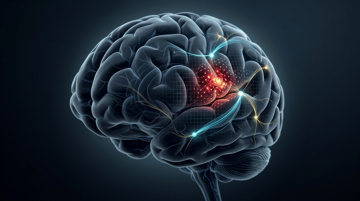
When an aneurysm on a brain artery ruptures and floods the space around the brain with blood, the condition known as aneurysmal subarachnoid hemorrhage, clinicians face one of the most unforgiving prognostic puzzles in medicine. Outcomes after aneurysmal subarachnoid hemorrhage vary widely between individuals, and current prognostic assessment mainly relies on clinical variables and CT findings. Doctors currently judge prognosis using clinical variables such as age, neurological grade and the amount of blood visible on a computed tomography scan, but these conventional models often lack biological interpretability, according to a new multicenter study published in Critical Care.
That study, drawn from the PROSAH-MPC registry and registered retrospectively as NCT05738083, set out to build and validate a prognostic model that fuses routine clinical information with quantitative features extracted from admission CT scans, a technique known as radiomics. The researchers then went a step further, performing exploratory cerebrospinal fluid proteomic characterization of the risk phenotypes their model defined, in an attempt to glimpse the biology that might underlie different recovery trajectories after the hemorrhage.
The cohort comprised 879 patients treated across four hospitals. Patients from the primary center were split into a training set of 637 and an internal validation cohort of 160, while patients from the three other centers formed an external validation cohort of 82. On each patient's admission CT, hemorrhagic lesions were automatically segmented using the validated BrainHemoAI platform, and radiomic features were extracted with PyRadiomics.
Three XGBoost models were then developed: one based on clinical variables alone, one on radiomic features alone, and one combining both. XGBoost models were developed to predict outcomes from the structured clinical and imaging data. To interpret the models, the team applied SHAP, or SHapley Additive exPlanations, which identifies the contribution of each input feature to the model's predictions.
The combined clinical-radiomics model achieved an area under the receiver operating characteristic curve, or AUC, of 0.892 in the internal test cohort and 0.820 in the external validation cohort. The AUC is a measure of the model's ability to discriminate between patients with different outcomes. Risk reclassification analyses suggested that the radiomic features may provide prognostic information that complements what clinical variables already capture.
The SHAP analysis identified radiomic heterogeneity, a measure of how irregularly the hemorrhage is textured and distributed within the segmented lesion, together with key clinical variables, as the major predictors driving the model’s output. In practical terms, the SHAP analysis identified radiomic heterogeneity, together with key clinical variables, as major predictors driving the model's output.
The second half of the study was more speculative by design. The researchers applied risk scores from the combined clinical-radiomics model to an independent prospective CSF proteomic cohort, dividing patients into model-defined high- and low-risk groups. They then compared the proteins detectable in the cerebrospinal fluid bathing the brain between these groups, using Gene Ontology and KEGG pathway enrichment, gene set enrichment analysis and protein-protein interaction network analyses to organize the resulting signals into biological themes.
This exploratory analysis identified candidate differentially expressed proteins and associated pathway signals that differed between the high- and low-risk groups defined by the combined model. In other words, patients the algorithm flagged as high risk carried a distinct molecular profile in their cerebrospinal fluid, though the analysis was exploratory and the identified signals require further validation. The authors frame this as hypothesis-generating work rather than proof of mechanism; the findings require validation before any of the flagged proteins or pathways can be considered therapeutic targets.
Even so, the study's architecture points to a plausible clinical workflow. A patient arrives with a suspected hemorrhage, the CT is acquired for diagnosis anyway, the automated segmentation tool BrainHemoAI delineates the blood, PyRadiomics converts the lesion into quantitative descriptors, and the model returns an interpretable risk estimate, with SHAP values showing which imaging and clinical features drove the number. Because the model was validated externally on patients from hospitals outside the primary center, its performance is less likely to reflect site-specific quirks in scanning protocols or patient mix, though the external cohort of 82 patients remains modest in size.
The authors conclude that the clinical-radiomics model enables accurate outcome prediction and provides exploratory biological insight into unfavorable recovery after aneurysmal subarachnoid hemorrhage, supporting refined risk stratification and potential targeted therapeutic development. For a condition in which every hour of early decision-making matters, a prognostic tool that is simultaneously accurate, externally validated and transparent about its reasoning, and that generates testable molecular hypotheses about why some patients fare worse than others, represents a meaningful step toward individualized care in one of neurocritical care’s most devastating emergencies.
Subject of Research: A clinical-radiomics machine learning model for predicting recovery after aneurysmal subarachnoid hemorrhage
Article Title: CT-based radiomics for interpretable outcome prediction after aneurysmal subarachnoid hemorrhage: a multicenter cohort study
Article References: Wu, Y., Hu, P., Yan, T., Xiao, Y., Qiu, Y., Sheng, J., Long, G., Wu, M., Zhang, Z., Cai, J., Wang, X., & Zhu, X. (2026). CT-based radiomics for interpretable outcome prediction after aneurysmal subarachnoid hemorrhage: a multicenter cohort study. Critical Care. https://doi.org/10.1186/s13054-026-06445-4
Image Credits: AI Generated
DOI: 10.1186/s13054-026-06445-4
Keywords: aneurysmal subarachnoid hemorrhage, radiomics, computed tomography, XGBoost, SHAP, machine learning, prognostic model, cerebrospinal fluid proteomics, risk stratification, multicenter cohort, BrainHemoAI, PyRadiomics
News Source: Cassandra Pierce. (November 4, 2026). CT Radiomics Model Predicts Outcomes After Bleeding Stroke Across Four Hospitals. Scienmag.