


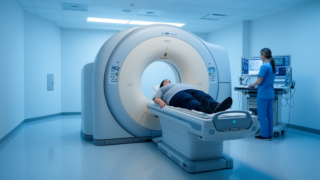
A PET/CT case report published in EJNMMI Reports describes the use of CT-less attenuation correction in a patient with one of the most extreme body habitus encountered in clinical imaging: a 34-year-old man weighing 264 kg and measuring 185 cm tall, giving a body mass index of 77. The report, authored by Twyla Bartel, Mickalia Johnston, Tracy Yarbrough, Kelly Harris, LaTaaron White and Stephen Lokitz, was published on 17 August 2026 as an open access article and documents how the technique performed when conventional CT-based attenuation correction would be expected to struggle most.
The patient was injected with 433 MBq (11.7 mCi) of the radiotracer [18F]FDG and underwent a routine PET/CT scan beginning 73 minutes after injection. The acquisition covered five bed positions at four minutes per bed. The CT component used 140 kVp, with a volumetric CT dose index of 17.895 mGy and a dose-length product of 2314.71 mGy·cm. Scanning was performed on the United Imaging Healthcare uMI Panorama PET/CT system, and the same raw data were reconstructed twice: once with conventional CT-based attenuation correction (CTAC) and once with CT-less attenuation correction, allowing a direct comparison of the two approaches in the same patient.
Both reconstructions used Deep Progressive Reconstruction, a vendor-provided AI-aided reconstruction method, with a user-defined strength parameter of 2 on a scale of 1 to 5, a 760 mm field of view, and a 256 × 256 matrix size. The comparison is therefore controlled for reconstruction parameters, isolating the effect of the attenuation correction method itself. The published figures include fused PET/CT slices, coronal slices and maximum intensity projections from the patient.
The most striking finding concerned respiration-induced PET/CT mismatch, which was particularly evident at the liver-lung interface on the fused images. CT-less attenuation correction, which derives the attenuation map directly from the PET data rather than from the CT, reduced these artifacts and improved the uniformity of liver uptake in the corrected images.
The report also notes that CT-less attenuation correction was applicable in this extremely large patient and reduced both PET/CT mismatch and CT truncation artifacts. Truncation is a persistent problem in large patients, because the CT field of view may not cover the full width of the body, leaving parts of the anatomy outside the attenuation map and introducing quantification errors. By removing the dependence on the CT for attenuation information, the technique sidesteps this limitation entirely, and it also has the potential to reduce radiation exposure by lessening reliance on the CT component of the examination.
According to the authors, the overall conclusion is that CT-less attenuation correction provides a viable alternative to conventional CTAC, with comparable image quality and quantification accuracy in challenging imaging conditions including extreme body habitus. The report places this single case in the context of recent advances in artificial intelligence and image reconstruction that have enabled CT-less attenuation correction methods for PET imaging, approaches that avoid CT-related artifacts such as truncation or misregistration while potentially reducing radiation exposure and maintaining quantitative accuracy.
High body mass index is highlighted as a setting where the technique is especially promising, since CT field-of-view limitations and degraded image quality can compromise attenuation maps and cause quantification errors precisely in the patients who are hardest to image. Early results with the uMI Panorama PET/CT system using in-house acquisition and reconstruction parameters had already suggested that CT-less attenuation correction produces PET images comparable to those obtained with CTAC, and this case extends that observation to a patient whose size pushes standard protocols to their limits.
As with any single-patient case report, the findings should be interpreted with appropriate caution: they demonstrate feasibility and artifact reduction in one individual rather than systematic performance across a patient population, and broader validation would be needed before drawing general conclusions about routine clinical use. Nevertheless, the case offers a concrete illustration of how AI-enabled reconstruction and PET-derived attenuation maps may expand the range of patients who can be imaged quantitatively with PET, including those whose body habitus has traditionally pushed CT-based workflows to their limits.
Subject of Research: CT-less attenuation correction for PET imaging in a patient with extreme obesity
Article Title: CT-less attenuation correction in a patient with BMI of 77
Article References: Bartel, T., Johnston, M., Yarbrough, T., Harris, K., White, L., & Lokitz, S. (2026). CT-less attenuation correction in a patient with BMI of 77. EJNMMI Reports, 10(1), Article 31. https://doi.org/10.1186/s41824-026-00314-7
Image Credits: AI Generated
DOI: 10.1186/s41824-026-00314-7
Keywords: PET/CT, attenuation correction, CT-less AC, BMI 77, FDG PET, truncation artifact, respiration mismatch, uMI Panorama, deep progressive reconstruction, image quality, radiation exposure, case report
News Source: Ophelia Keating. (October 29, 2026). CT-less PET attenuation correction proves viable in a patient with BMI 77. Scienmag.



