Researchers have established a correlative workflow that combines X-ray microscopy with transmission electron microscopy to analyze the intracellular ultrastructure of zebrafish neuromasts. In a technical report published in Applied Microscopy, investigators Sun-Yeong Gwon, MinKi Choi, and Ji Young Mun demonstrated that pre-screening resin-embedded tissue blocks with non-destructive three-dimensional X-ray imaging streamlined subsequent ultra-structural examination while reducing specimen loss and trimming errors.
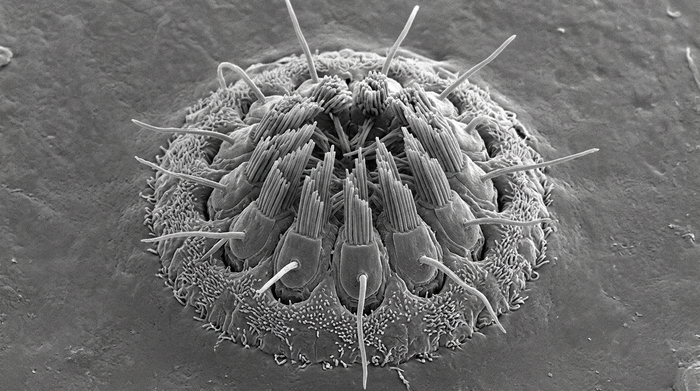
The zebrafish neuromast is a specialized mechanosensory organ that detects water flow through ciliary motion. Located laterally between the dorsal and ventral sides of the zebrafish, the neuromast measures approximately 20 to 30 micrometers in diameter and extends to a depth of 30 to 50 micrometers from the skin surface. Structurally, the organ consists of hair cells, supporting cells, and mantle cells arranged along the animal’s lateral line.
Investigating these cellular architectures with conventional transmission electron microscopy presents persistent technical challenges. Although transmission electron microscopy provides high spatial resolution for cellular ultrastructure, standard protocols require extensive trimming and trial-and-error semi-thin sectioning using an ultramicrotome. Because heavy osmium staining makes targets difficult to distinguish under light microscopy, technicians traditionally cut serial semi-thin slices at intervals of ten to twenty sections and apply toluidine blue stains to verify specimen depth and orientation.
To overcome this bottleneck, the researchers integrated non-destructive X-ray microscopy prior to physical trimming. Zebrafish tissue specimens obtained from KRIBB were initially fixed with 2 percent paraformaldehyde and 2.5 percent glutaraldehyde. Following chemical fixation, samples were processed according to the National Center for Microscopy and Imaging Research protocols for three-dimensional electron microscopy, which included ferrocyanide-reduced osmium tetroxide incubation, thiocarbohydrazide-osmium ligation staining, uranyl acetate, and lead aspartate contrasting before dehydration and embedding in hard EMbed 812 resin.
Rather than relying immediately on mechanical sectioning, the intact resin blocks were scanned using a Carl Zeiss Xradia 620 Versa system equipped with a 20× objective lens, operating at an acceleration voltage of 80 kilovolts and a power of 10 watts. Imaging was performed in fan mode over a 180-degree rotation angle with an LE4 filter, collecting 4,501 projection images at one-second exposures for a total scan time of roughly 4.5 hours, yielding a reconstructed voxel size of 0.6 micrometers.
Reconstruction of the X-ray projection datasets was executed with DeepRecon Pro, a machine learning algorithm designed for noise reduction and image quality enhancement. Using Dragonfly software, the team visualized the three-dimensional block geometry across multiple planar and orthogonal perspectives, generating cross-sectional views along the X, Y, and Z axes to map internal anatomical landmarks.
The resulting three-dimensional renderings allowed direct identification of muscle tissue, neurons, notochord, and yolk sac within the opaque resin. The neuromast appeared noticeably brighter than adjacent tissue structures, exhibiting a distinctive morphology. Beneath the sensory organ, the researchers identified a bright region likely representing the myelinated lateral line nerve, consistent with the higher X-ray attenuation typical of myelin.
The three-dimensional spatial coordinates established by X-ray microscopy provided precise measurements of target depth, tilt, and surface distance. While ideal resin-embedded preparations orient parallel to the anterior block face, most actual tissues are embedded at an angle. Software-guided rotation and section previews permitted researchers to adjust the trimming angle accurately, minimizing the risk of sectioning past the organ or slicing at suboptimal angles.
Following software localization, targeted trimming removed excess resin, and semi-thin 500-nanometer sections confirmed that predicted anatomical coordinates aligned closely with toluidine blue histology. Subsequent ultrathin sectioning between 70 and 100 nanometers was carried out on an ultramicrotome, and the resulting sections were collected on Formvar-coated one-hole grids for transmission electron microscopy at an operating voltage of 120 kilovolts.
Transmission electron microscopy verified that the correlative workflow preserved fine cellular features. High-resolution micrographs analyzed with DigitalMicrograph and ImageJ Fiji resolved hair cells, supporting cells, cell-to-cell contacts, nuclei, and mitochondrial morphology. The authors concluded that incorporating X-ray microscopy previews into volume electron microscopy workflows offers a reproducible strategy to optimize orientation accuracy and conserve delicate biological targets.
The historical emergence of X-ray microscopy traces back to the period following Röntgen’s discovery of X-rays, which catalyzed the development of projection microscopes capable of interrogating biological specimens at nanometer resolution. Over subsequent decades, the modality expanded considerably as researchers combined X-ray optics with spectroscopic techniques. Within biological laboratories, transmission X-ray microscopy established a method for obtaining high-resolution structural information through single full-field projections. When integrated with computed tomography, these systems generate complete three-dimensional reconstructions of intact tissues, providing anatomical context across thick biological samples without requiring preliminary mechanical slicing.
Specialized variations of X-ray imaging have further expanded investigations into subcellular organization and biochemical distributions. For instance, water-window X-ray microscopes coupled with wide-field transmission microscopy permit the simultaneous acquisition of both two-dimensional and three-dimensional structural data from biological specimens. In parallel, advanced micro-tomography techniques, including scanning transmission X-ray microscopy and X-ray fluorescence microspectroscopy, enable the precise mapping of elemental distributions across internal tissue environments. These spectroscopic approaches have proven particularly valuable for elucidating intracellular physiological mechanisms and tracking nanoparticle interactions within cells. By delivering non-destructive spatial and chemical data across relatively large volumes, X-ray technologies bridge the observational gap between gross anatomical structures and ultra-fine cellular architectures.
Although scanning electron microscopy has achieved substantial improvements in spatial resolution, many biological researchers continue to prefer transmission electron microscopy due to its superior resolution for cellular ultrastructure and diverse analytical applications. Nonetheless, traditional transmission electron microscopy workflows have long been constrained by mechanical sample preparation bottlenecks. Prior efforts to mitigate these issues combined X-ray microscopy with serial block-face scanning electron microscopy, which demonstrated substantial enhancements in tracking regions of interest and improving targeting efficiency. Extending a correlative workflow to transmission electron microscopy pairs the broad, non-destructive volumetric capabilities of X-ray imaging with the ultimate high-resolution visualization of intracellular organelles and membrane contacts.
To achieve the heavy-metal contrast required for volume imaging, the researchers implemented precise wet-laboratory procedures following primary aldehyde fixation. Excised zebrafish tissues were first washed three times in cold cacodylate buffer for five minutes each. Next, the samples underwent a one-hour incubation on ice in a buffered heavy-metal cocktail combining equal parts of 3 percent potassium ferrocyanide in 0.3 molar cacodylate buffer and 4 percent aqueous osmium tetroxide, yielding a final concentration of 1.5 percent potassium ferrocyanide and 2 percent osmium tetroxide in 0.15 molar cacodylate buffer. In a parallel preparation step, 0.1 grams of thiocarbohydrazide was dissolved in ten milliliters of double-distilled water, heated in an oven at 60 degrees Celsius for one hour with gentle swirling every ten minutes, and passed through a 0.22-micrometer syringe filter. After the initial osmium treatment, the tissues were rinsed three times in double-distilled water at room temperature for five minutes per rinse, incubated in the freshly filtered thiocarbohydrazide solution for twenty minutes at room temperature, rinsed again three times in double-distilled water for five minutes each, and placed in 2 percent aqueous osmium tetroxide for thirty minutes at room temperature.
The contrasting sequence concluded with overnight incubation in 1 percent uranyl acetate at 4 degrees Celsius following three room-temperature rinses in double-distilled water. The tissue pieces were washed again in double-distilled water, placed into lead aspartate solution in an oven at 60 degrees Celsius for thirty minutes, and rinsed. Dehydration proceeded through ascending grades of ice-cold ethanol, comprising 20, 50, 70, 90, and two changes of 100 percent anhydrous ethanol for ten minutes each, before samples were transferred into two ten-minute baths of anhydrous ice-cold 100 percent acetone at room temperature. Infiltration was carried out using hard EMbed 812 resin formulated by weight and mixed with acetone by concentration. Tissues were immersed progressively in 25 percent resin for two hours, 50 percent resin for two hours, and 75 percent resin for two hours, followed by 100 percent resin overnight and fresh resin for two hours. Finally, specimens were embedded in silicone molds and cured in an oven at 60 degrees Celsius for 48 hours.
To navigate the target organ accurately within the block geometry, the researchers evaluated structural schematics comparing lateral and dorsal orientations of the zebrafish. These diagrams used circular pink boundaries to designate the cross-sectional area of the neuromast, while sky-blue and blue trimming boxes indicated the sampling boundaries, with the blue perimeter extended dorsally to capture the true depth of the organ within the tissue. During volumetric scanning on the Xradia 620 Versa instrument installed at the Zeiss Innovation Center in Dongtan, Korea, the rotation tool within Dragonfly software permitted examination of the anterior specimen from front, right, left, top, and bottom perspectives, clearly displaying the outer resin boundaries as grey lines. Within orthogonal cross-sections, distinct anatomical landmarks were documented using specific visual indicators, including orange arrowheads for muscle tissue, white arrowheads for neurons, grey arrowheads for the notochord, and pink arrowheads for the yolk sac. Electron microscopy validation incorporated large stitched overviews at 1,700-times magnification, resolving the nucleus marked by yellow arrows, mitochondria marked by green arrows, and cell-to-cell interactions marked by blue arrows. The authors concluded that this workflow assists volume electron microscopy across diverse biological tissues, with datasets available from the corresponding author upon reasonable request.
Subject of Research: Correlative X-ray and transmission electron microscopy of zebrafish neuromasts
Article Title: Correlative x-ray microscopy and transmission electron microscopy for ultrastructural analysis of zebrafish tissue
Article References: Gwon, S.-Y., Choi, M., & Mun, J. Y. (2025). Correlative x-ray microscopy and transmission electron microscopy for ultrastructural analysis of zebrafish tissue. Applied Microscopy, 55(1), Article 15. https://doi.org/10.1186/s42649-025-00120-8
Image Credits: AI Generated
DOI: 10.1186/s42649-025-00120-8
Keywords: X-ray microscopy, transmission electron microscopy, correlative microscopy, zebrafish, neuromast, ultrastructure, sample preparation, volume electron microscopy, Correlative, x-ray, microscopy, transmission
News Source: Denise Maddox. (November 7, 2026). Correlative X-Ray and Electron Microscopy Resolves Zebrafish Neuromast Ultrastructure. Scienmag.