Researchers have introduced a measurement technique called continuous mass photometry that enables hour-long, mass-resolved observation of individual biomolecules tethered to supported lipid bilayers, together with their complexes and interactions, with up to sub-kilodalton mass precision and millisecond temporal resolution. The work, published as a Brief Communication in Nature Methods by a team at the University of Oxford led by Philipp Kukura, presents the approach as a quantitative, time-resolved, single-molecule measurement modality for studying complex biomolecular mechanisms in action. Rather than recording a single mass snapshot per molecule, the technique holds each molecule within a defined observation area and follows its mass continuously as binding events occur.
The motivation set out in the abstract is that the dynamic choreography of biomolecular interactions underpins the processes of life, but its direct observation remains challenging. Continuous mass photometry addresses this by keeping the molecules of interest in place on a supported lipid bilayer, a surface to which proteins can be tethered, and then repeatedly interrogating the same individual particles with a scanning laser. Because the tethered molecules stay within the observation region, the instrument can build up a continuous mass trajectory for each one over periods approaching an hour, rather than losing each molecule after a single landing event.
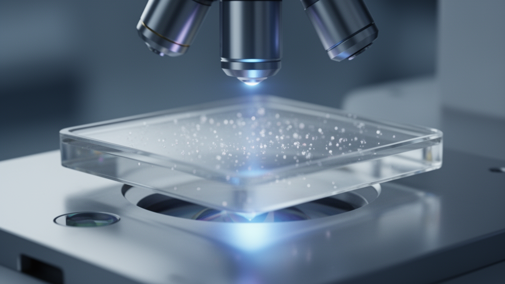
The optical setup is a home-built mass photometer equipped with a 638-nanometer red diode laser from Lasertack and a Blackfly 17S7M-C FLIR camera, with acousto-optic deflector scanning controlled by a Digilent Analog Discovery 2 device and operation handled through a home-built LabVIEW script. Measurements were performed in a field of view of approximately 6 by 6 micrometers, centered on a trap roughly 3.5 micrometers in diameter. Images were acquired with an exposure time of 1,700 microseconds at frame rates that varied by experiment type: 497 hertz for the long tracking experiments, 250 hertz for the step change experiments and 10 hertz for the transient binding experiments. For the step change and transient binding measurements, the acousto-optic deflector scanning was pulsed to position the beam further from the trap when the camera was not exposed.
Data processing relied on an in-house Python script adjusted for single-trap analysis. Proteins were detected using a Laplacian of Gaussian threshold filter applied to a median-processed mass photometry movie, a step that reveals the much smaller protein signal, which amounts to roughly 0.5 percent of the raw image. Detected proteins were then fitted to an analytical point spread function calibrated to the home-built setup to determine the protein contrast, and the fitted events were filtered for single-protein analysis. The starting mass in each trajectory was set as the mean mass obtained from a conventional landing assay. Results were presented as a running median of the raw fitted event masses or as averaged mass values of consecutive time windows. For step detection, the team implemented a step detection algorithm applying a maximum threshold of ten steps per trajectory and minimum step size thresholds of 100 kilodaltons for antibodies, 30 kilodaltons for Fabs and 8 kilodaltons for nanobodies. The transient binding data were time-averaged to 0.2 hertz and fitted to a two-state hidden Markov model.
The sample preparation underlying each measurement is exacting. Liposomes were prepared by dissolving POPC, DOPE-PEG550 and DGS-NTA(Ni) lipids in chloroform and mixing them at a 97:2:1 molar ratio, corresponding to a total lipid concentration of 500 micromolar in 0.5 milliliters of buffer. The mixture was dried under a constant nitrogen stream via rotary evaporation and further dried under vacuum for one hour at room temperature, then rehydrated with 500 microliters of HKS-150 buffer containing 20 millimolar HEPES at pH 7.4 and 150 millimolar KCl. After incubation at 50 degrees Celsius for one hour and brief vortexing, the liposome mixture was stored overnight at room temperature, sonicated in an ice-water bath using a 3-millimeter probe at 25 percent amplitude with a one-second-on, three-second-off cycle for ten minutes of active sonication, and then spun at 21,130 times gravity for 30 minutes at 4 degrees Celsius. The supernatant was stored at 4 degrees Celsius and used within 30 days.
Patterned supported lipid bilayers were prepared on glass coverslips that were cleaned by sonication in Milli-Q water and then in a 50:50 Milli-Q water and isopropanol solution, dried with nitrogen, and plasma cleaned at 40 percent power for five minutes. The coverslips were incubated with a 2-microgram-per-milliliter solution of PLL(20)-g[3.5]-PEG(2) for 30 minutes, rinsed, and then exposed to deep ultraviolet light in a mask aligner for 60 minutes using a home-made photolithography mask to create the patterned bilayer regions. The patterned coverslips could be stored at minus 20 degrees Celsius for up to three months before use. Before each experiment, a coverslip was brought to room temperature and exposed to 25 microliters of fusion buffer, containing 20 millimolar HEPES at pH 7.4, 150 millimolar KCl and 2 millimolar MgCl2, together with 5 microliters of liposome solution in a silicone gasket. Bilayer formation, monitored by mass photometry, took around five to ten minutes, after which the surfaces were washed five times with phosphate-buffered saline, blocked with 0.1 milligram per milliliter PLL-PEG for 30 minutes unless otherwise noted, and washed again ten times by solvent volume replacement.
To demonstrate the technique, the researchers applied it to SARS-CoV-2 spike trimers. Spike was added at a concentration of 5 nanomolar in solution for capture and washed away within 30 seconds, yielding a capturing occupancy of zero to two particles per confined supported lipid bilayer. In the long tracking experiments, the supported lipid bilayers were not blocked before protein addition, and the trap was illuminated continuously at a 497-hertz frame rate with the 1,700-microsecond exposure time, allowing individual tethered spike trimers to be followed over extended durations. In the step change experiments, after a three-minute baseline period with no interaction partner in order to establish a baseline mass for each trimer, an antibody, Fab or nanobody was added at the three-minute mark. Antibodies were added to a final concentration of 100 nanomolar by a 0.75:99 dilution from a 13.3 micromolar stock, and the solution was mixed by pipette every five minutes. Fabs and nanobodies were each added to a final concentration of 100 nanomolar by a 10:90 dilution from a 1 micromolar stock, with an additional 10 microliters of stock added every five minutes to facilitate binding within the measurement period.
The transient binding experiments captured interactions that do not persist as permanent mass steps. Spike trimers were again added at 5 nanomolar and washed within 30 seconds to reach a capturing occupancy of zero to two particles per confined bilayer, and the trap was illuminated with pulsed illumination at a 10-hertz frame rate. After the same three-minute baseline, 210 nanomolar of mACE2 was added while the spike was continuously measured. The resulting data were time-averaged to 0.2 hertz and fitted to a two-state hidden Markov model, allowing the bound and unbound states of individual tethered spike trimers to be distinguished as the interaction partner repeatedly attached and released during the recording.
Alongside the continuous measurements, the team performed landing assay mass photometry on a commercial Refeyn TwoMP instrument at the regular field of view of 10.9 by 4.3 micrometers and 498 hertz, on slides cleaned by consecutive sonication in Milli-Q water followed by the 50:50 water and isopropanol solution. Interaction mixtures were incubated at equimolar concentrations of 25 nanomolar for 10 minutes and then measured as a 1:1 dilution with phosphate-buffered saline in the mass photometry gasket for 60 seconds. Frames were averaged to navg equal to 15, corresponding to 33.2 hertz, using the advanced software settings in DiscoverMP software version 2024 R1, and results are reported as the mean value of three technical replicates. These landing assay measurements provided the mean starting masses used to anchor the continuous mass trajectories.
The raw data required to reproduce the main figures, together with the Python software and Jupyter notebooks used for the analysis, have been deposited in the University of Oxford Research Archive, allowing other laboratories to reproduce the processing pipeline. As presented by the authors, continuous mass photometry extends label-free single-molecule mass measurement into a time-resolved regime: individual tethered biomolecules can be observed for up to an hour, their binding partners resolved as discrete mass steps down to the nanobody-scale thresholds used in the analysis, and short-lived interactions separated into bound and unbound states through hidden Markov modeling. The published demonstrations are limited to the SARS-CoV-2 spike system and the specific antibodies, Fabs, nanobodies and mACE2 used in the study, and the home-built nature of the optical setup means that reproducing the technique requires comparable instrumentation and analysis expertise.
Subject of Research: Continuous mass photometry for time-resolved single-molecule mass measurement of biomolecular interactions
Article Title: Continuous mass photometry by single-molecule trapping
Article References: Loewenthal, D., Asor, R., Thorpe, S., Peters, J. S., Zouboulis, K. C., Kaplan, M., Thiele, J. C., Bardzil, J., Helmi, S., Burnap, S. A., Tan, T. K., Struwe, W. B., Benesch, J. L. P., & Kukura, P. (2026). Continuous mass photometry by single-molecule trapping. Nature Methods. https://doi.org/10.1038/s41592-026-03252-1
Image Credits: AI Generated
DOI: 10.1038/s41592-026-03252-1
Keywords: mass photometry, single-molecule biophysics, supported lipid bilayer, SARS-CoV-2 spike, antibody binding, nanobodies, mACE2, label-free detection, hidden Markov model, optical setup, Nature Methods, University of Oxford
News Source: Drew Townsend. (October 26, 2026). Continuous mass photometry tracks single biomolecules for hours. Scienmag.