• HOME
  • NEWS
  • EXPLORE
    • CAREER
      • Companies
      • Jobs
    • EVENTS
    • iGEM
      • News
      • Team
    • PHOTOS
    • VIDEO
    • WIKI
  • BLOG
  • COMMUNITY
    • FACEBOOK
    • INSTAGRAM
    • TWITTER
Tuesday, August 4, 2026
BIOENGINEER.ORG
No Result
View All Result
  • Login
  • HOME
  • NEWS
  • EXPLORE
    • CAREER
      • Companies
      • Jobs
        • Lecturer
        • PhD Studentship
        • Postdoc
        • Research Assistant
    • EVENTS
    • iGEM
      • News
      • Team
    • PHOTOS
    • VIDEO
    • WIKI
  • BLOG
  • COMMUNITY
    • FACEBOOK
    • INSTAGRAM
    • TWITTER
  • HOME
  • NEWS
  • EXPLORE
    • CAREER
      • Companies
      • Jobs
        • Lecturer
        • PhD Studentship
        • Postdoc
        • Research Assistant
    • EVENTS
    • iGEM
      • News
      • Team
    • PHOTOS
    • VIDEO
    • WIKI
  • BLOG
  • COMMUNITY
    • FACEBOOK
    • INSTAGRAM
    • TWITTER
No Result
View All Result
Bioengineer.org
No Result
View All Result
Home NEWS Science News Health

Constructing new tissue shapes with light

Bioengineer by Bioengineer
June 18, 2018
in Health
Reading Time: 2 mins read
0
Share on FacebookShare on TwitterShare on LinkedinShare on RedditShare on Telegram
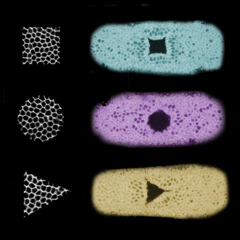
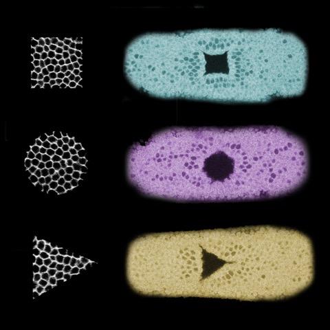
IMAGE

Credit: Stefano De Renzis, EMBL

Constructing biological tissues, such as skin, muscle, or bone, in customized shapes is now one step closer. Researchers at EMBL have succeeded in guiding the folding and thus shape of tissues with optogenetics: a technique to control protein activity with light. Nature Communications publishes their results, with implications for regenerative medicine, on 18 June.

The changing of tissue shapes in an embryo is essential for healthy development. Stefano De Renzis and his group members at EMBL are interested in the mechanisms behind these shape transitions, also called morphogenesis. They use optogenetics – a technique providing precise light-mediated control of protein activity – to study changes in tissue shapes.

Uncoupling the link between shape and function

In the current paper, Emiliano Izquierdo, Theresa Quinkler, and Stefano De Renzis used optogenetics to reconstruct epithelial folding: a fundamental process during development, where cells move inwards and fold into the embryo, eventually giving rise to internal tissues like muscles, for example. Remarkably, they achieved this in cells that normally do not undergo this process. De Renzis, who led the study: "We've uncoupled the link between the shape and function of a cell. This allows us to, for the first time, built tissues in certain shape without affecting the cell's expertise."

Image 1. Three examples of the tissue shapes the team created. The black and white square, circle and triangle on the left correspond to the cells that were illuminated. On the right, three fruit fly embryos are shown in cyan, magenta and yellow, demonstrating how the illuminated cells folded inwards after the light-activation.

Precise control

"The great thing about using optogenetics to guide morphogenesis is that it is a very precise technique", says Emiliano Izquierdo, first author of the study. "We were able to define various shapes, and by alternating the timing and strength of illumination, we could control how far the cells folded inwards."

Image 2. Example of optogenetics-guided tissue folding. The top image shows an embryo 10 minutes after illumination , and the bottom one 13 minutes afterwards: light-activated cells have folded inwards and thus moved downwards, creating a furrow.

From fruit fly to the clinic?

The research was done in developing fruit flies, but since epithelial folding is a conserved process across evolution, De Renzis expects these methods to also be applicable in other organisms and ex vivo stem cell culture systems. In that case, optogenetics could be an ideal technique for reconstructing and directing tissue development, which could be used to (re)build artificial tissues in regenerative medicine.

###

Video about optogenetics and how the technique is used at EMBL.

Media Contact

Iris Kruijen
[email protected]
49-622-138-78443
@EMBL

http://www.embl.org

Related Journal Article

http://dx.doi.org/10.1038/s41467-018-04754-z

Share12Tweet8Share2ShareShareShare2

Related Posts

Chronic Disease Burden Links Frailty, Sleep, and Life Satisfaction in Older Adults

August 4, 2026

White Matter Disorders Link Transcription, RNA Processing, and Translation

August 4, 2026

Routine Military Testosterone Screening Could Advance Evidence-Based Men’s Health

August 3, 2026

How the Outer Kinetochore Is Built and Functions

August 3, 2026
Please login to join discussion

POPULAR NEWS

  • Chronic Disease Burden Links Frailty, Sleep, and Life Satisfaction in Older Adults

    29 shares
    Share 12 Tweet 7
  • White Matter Disorders Link Transcription, RNA Processing, and Translation

    29 shares
    Share 12 Tweet 7
  • Routine Military Testosterone Screening Could Advance Evidence-Based Men’s Health

    29 shares
    Share 12 Tweet 7
  • How the Outer Kinetochore Is Built and Functions

    29 shares
    Share 12 Tweet 7

About

We bring you the latest biotechnology news from best research centers and universities around the world. Check our website.

Follow us

Recent News

Chronic Disease Burden Links Frailty, Sleep, and Life Satisfaction in Older Adults

White Matter Disorders Link Transcription, RNA Processing, and Translation

Routine Military Testosterone Screening Could Advance Evidence-Based Men’s Health

Subscribe to Blog via Email

Enter your email address to subscribe to this blog and receive notifications of new posts by email.

Join 86 other subscribers
  • Contact Us

Bioengineer.org © Copyright 2023 All Rights Reserved.

Welcome Back!

Login to your account below

Forgotten Password?

Retrieve your password

Please enter your username or email address to reset your password.

Log In
No Result
View All Result
  • Homepages
    • Home Page 1
    • Home Page 2
  • News
  • National
  • Business
  • Health
  • Lifestyle
  • Science

Bioengineer.org © Copyright 2023 All Rights Reserved.