The third iteration of the Cnidarian Model Systems Meeting, known as Cnidofest, took place from August 14 to 17, 2024, at Lehigh University in Bethlehem, Pennsylvania. This gathering brought together researchers from 11 countries to discuss advancements in cnidarian biology, a field that has seen significant growth in recent years. The meeting featured 48 oral presentations, with 90% delivered by trainees and the remaining 10% by principal investigators. A total of 37 posters were also presented across two sessions, highlighting the broad scope of research currently being conducted in this area.
A notable trend observed at Cnidofest 2024 was the increasing parity between cnidarian model systems and classical laboratory models such as Drosophila and vertebrates. Modern genomic, genetic, and molecular tools are now routinely applied to cnidarian research, allowing for more detailed and precise studies. This shift has enhanced the community’s ability to gain fundamental insights into animal biology that are often difficult to achieve in complex bilaterian model systems. The integration of cnidarian and bilaterian models provides researchers with a broader toolkit for addressing specific biological questions.
The meeting included three technology talks, a hallmark of Cnidofest events, designed to introduce emerging and cutting-edge technologies to the cnidarian research community. Blair Benham-Pyle from Baylor College of Medicine discussed spatial transcriptomics, a technique that combines single-cell RNA sequencing with fluorescence-activated cell sorting to map cellular and molecular microenvironments. This approach could advance cnidarian regeneration research by revealing the molecular pathways and signaling networks that drive tissue repair and stem cell activation.
Yongxin Zhao from Carnegie Mellon University presented his group’s ‘Magnify’ protocol for next-generation expansion microscopy. This technique allows for high-resolution nanoimaging using standard microscopes by enlarging biological samples embedded in a crosslinked hydrogel. ‘Magnify’ can expand samples up to 11 times, achieving around 25-nm resolution with conventional microscopes and up to 15-nm resolution with super-resolution techniques. This is particularly valuable for studying the complex tissue architectures of cnidarians, such as the intricate arrangement of neurons, cnidocytes, and epithelial cells.
Hang Lu from the Georgia Institute of Technology discussed the automation of optogenetic manipulation for small invertebrates. Her group’s research focuses on developing microfluidic devices and biomedical microelectromechanical systems to explore neuroscience, genetics, and biotechnology. Lu’s approach provides a method to predict behavioral responses based on sensory neuron activity, which is highly adaptable for a range of small invertebrates. These lab-on-a-chip tools offer large-scale quantitative data through miniaturized devices that leverage micro and nano-scale phenomena.
Oral presentations at the meeting covered a wide range of topics, including regeneration, developmental innovation, genomics, symbiosis, cell biology, physiology, and neurobiology. Ben Cox from the Juliano Lab at UC Davis discussed complex tissue remodeling during Hydra vulgaris head regeneration. Cox found that matrix metalloproteinases are required for collagen degradation during head regeneration and demonstrated that interstitial stem cells migrate from host tissue to populate the grafted head of the regenerate.
Marylene Bonvin from the Tsiairis Lab at the Friedrich Miescher Institute for Biomedical Research investigated the role of metabolic signaling in cell fate decisions of Hydra vulgaris tissue patterning. By overexpressing wnt, Bonvin observed a graded glycolytic activity along the body axis, implicating hexosamine pathways and O-glycosylation for efficient signaling. Iris Y Juanico, also from the Juliano Lab, explored the gene regulatory network of regenerating Hydra vulgaris, finding that wnt9/10C and wntless transcription is dependent on ERK signaling, while wnt3 transcription is not.
Clara Nuninger from the Tsiairis Lab discussed the mechanism of morphallaxis in Hydra vulgaris regeneration. Using single-cell RNA sequencing, Nuninger found that cells at the regenerating extremities recreate their first missing neighbors and progressively acquire their correct axial positions. Sera Weever, also from the Tsiairis Lab, observed positive feedback between tissue stretching and Wnt3 signaling during Hydra vulgaris regeneration, supported by a mathematical model that does not require the presence of a long-range diffusible inhibitor.
Justin Waletich from the Schnitzler Lab at UF Whitney Marine Lab identified eight new markers of the adult Hydractinia symbiolongicarpus pluripotent stem cell population from a single-cell atlas. The atlas revealed one ‘germ-like’ and one ‘somatic-like’ i-cell cluster, with specific genes expressed in each. Yu-ichiro Nakajima from the University of Tokyo presented on the regenerating tentacles of the hydrozoan jellyfish, Cladonema pacificum, finding that cell proliferation is directly controlled by the ERK/MAPK signaling pathway, while proliferation of resident stem cells is controlled by Notch signaling.
Research on developmental innovation included work by Kennedy Bolstad from the Babonis Lab at Cornell University on tissue remodeling in the staurozoan Haliclystus sanjuanensis. Bolstad confirmed a shift in investment from juvenile tentacles to adult anchors and demonstrated that it takes 48 hours to replace fired cnidocytes in the feeding tentacles. Bailey Steinworth from the Martindale Lab at the University of Florida explored the redeployment of embryonic Hox genes during asexual reproduction in the scyphozoan Cassiopea xamachana, revealing similar patterns in both developing embryos and asexual buds.
Matthew Travert from the Cartwright Lab at Kansas University analyzed RNAseq data from four medusozoan species to identify genes specific to different life stages. He identified 24 ANTP-class homeobox transcripts shared across all four species, with 33% showing medusa-specific expression. Sarah Arnold from the Babonis Lab pioneered the Northern Star Coral, Astrangia poculata, as a model for studying cell type diversification, showing that both life stages possess a broader diversity of cnidocytes than the sea anemone Nematostella vectensis.
Recent advances in cnidarian genomics were highlighted by Namrata Ahuja from the Dunn Lab at Yale University, who performed single-cell RNA sequencing on two siphonophore species to identify cell types. Alberto Rivera from the Baxevanis Lab at NIH used genome annotation software to investigate the evolutionary history of allorecognition in cnidarians, finding structural conservation between cnidarian allorecognition proteins and bilaterian immune system major histocompatibility complex proteins.
The meeting also covered cnidarian symbiosis, with Shumpei Maruyama from the Cleves Lab at Carnegie Science isolating membrane-bound compartments containing symbiotic Symbiodiniaceae spp. from the sea anemone Exaiptasia diaphana. Miranda Gibson from the Titus Lab at the University of Alabama used ddRADseq and ultra-conserved element phylogenomics to identify diversity in the tropical clownfish-hosting sea anemone Radianthus magnifica. Sophie Macvittie from the Sogin Lab at the University of California, Merced, explored the impacts of antibiotic exposure on the facultative symbiotic sea anemone E. diaphana, finding that microbial load recovered fully after three weeks.
Cell biology research included work by Sue Xu from the Schnitzler Lab on the Hippo pathway in Hydractinia symbiolongicarpus, finding that yki has a role in promoting budding and regeneration. David Ehrens from the Traylor-Knowles Lab at the University of Miami identified thraustochytrids as decomposers in cnidarians’ mucus and tissues and proposed a protocol to eliminate them from coral tissue samples. Ines Fournon Berodia from the Steinmetz Lab discussed epithelial remodeling in response to food availability in Nematostella vectensis, identifying rosette-like epithelial structures indicative of cell extrusion.
Physiology research included work by Daria Aleshkina from the Moran Lab at Hebrew University on the roles of RNA-dependent RNA polymerases in the antiviral response of Nematostella vectensis. Janki Bhalodi from the Reitzel Lab at UNC Charlotte identified the genome-wide distribution patterns of heat shock factor binding sites in various cnidarians. Benjamin Glass from the Barott Lab at the University of Pennsylvania discussed the susceptibility of three species to an experimentally simulated hypoxia event, finding a negative impact on early life stages.
Neurobiology research was a significant focus, with Wataru Yamamoto from the Yuste Lab at Columbia University exploring neural mechanisms driving osmoregulation in Hydra vulgaris. Ehsan Sakib from the Bosch Lab at Christian Albrechts University used asexual development in Hydra vulgaris to understand how a new nerve net develops. Christopher Noack from the Bosch Lab developed a methodology to investigate the formation of neural networks de novo in the developing Hydra vulgaris embryo. Hannah Justin from the Reitzel Lab constructed a platform to automate the delivery of mechanical stimuli to Nematostella vectensis to assess its capacity to habituate to repeated stimuli.
Developmental research included work by MingHe Cheng from the Layden Lab at Lehigh University on neuronal subtype identity patterning in Nematostella vectensis, finding support for at least two waves of neurogenesis. Benjamin Danladi from the Layden Lab presented work on the role of the transcription factor NvfoxD-like2 in Nematostella vectensis cnidogenesis. Bert Hobmayer from the University of Innsbruck presented work on the function of Myc in Hydra vulgaris, suggesting that Myc2 acts as a stem cell maintenance factor. Hannah Morris Little from the Juliano Lab explored Wnt signaling in Hydra vulgaris, finding that Wnt signaling regulates distinct transcriptional programs depending on cellular context.
Ulrich Technau from the University of Vienna presented work showing that Notch signaling regulates endodermal specification in Nematostella vectensis, which may be a shared ancestral feature with bilaterians. Layla Al-Shaer from the Layden Lab produced one of the first differential expression analyses of both male and female Nematostella vectensis peri-induction of their spawning cycles. Mako Takahashi from the Kumano Lab at Tohoku University investigated the temporal landscape of female germline cell determination in Cladonema pacificum. Sergio Stampar from São Paulo State University conducted ultrastructural analysis of reproductive tissues from 18 Ceriantharia species, revealing diverse reproductive strategies.
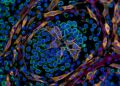
Yamini Ravichandran from the Roux Lab at Université de Genève concluded the meeting with a presentation on the function of topological defects in actin organization during head regeneration in Hydra vulgaris. Ravichandran presented evidence that topological defects in the actin cytoskeleton participate in initiating regeneration from small tissue fragments. These findings are reinforced by the presence of similar topological defects in other biological systems, supporting the idea that actin-based topological defects are a conserved mechanism contributing to morphogenesis across diverse animal systems.
Subject of Research: Advancements in cnidarian biology research and the integration of modern genomic and molecular tools at Cnidofest 2024
Article Title: Discoveries and innovations in cnidarian biology at Cnidofest 2024
Article References: Leach, W. B., Babonis, L., Juliano, C. E., Nakanishi, N., Schnitzler, C. E., Steinmetz, P. R. H., & Layden, M. J. (2025). Discoveries and innovations in cnidarian biology at Cnidofest 2024. EvoDevo, 16(1), Article 9. https://doi.org/10.1186/s13227-025-00247-5
Image Credits: AI Generated
DOI: 10.1186/s13227-025-00247-5
Keywords: cnidarian biology, Cnidofest 2024, genomic tools, molecular tools, regeneration, neurobiology, developmental biology, symbiosis, cell biology, physiology, Discoveries, innovations
News Source: Drew Townsend. (November 9, 2026). Cnidofest 2024 Highlights New Cnidarian Research Tools. Scienmag.