


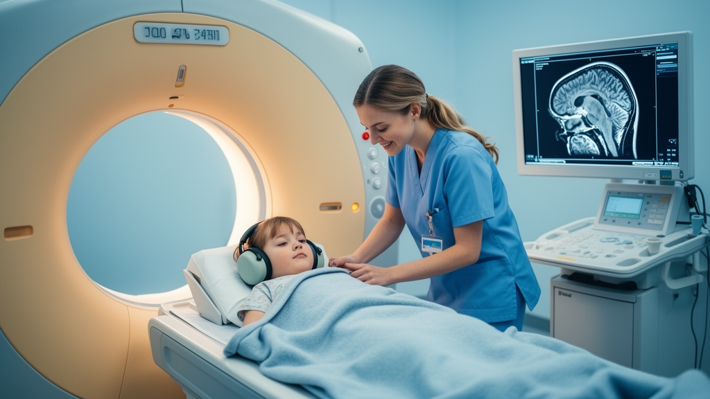
A carefully designed diffusion-weighted MRI study of children with migraine has delivered a result that is as notable for what it did not find as for what it hints at. Researchers comparing apparent diffusion coefficient, or ADC, values across seven brain regions in 48 children with migraine and 48 non-migraine clinical controls found that not a single regional difference remained statistically significant once the analysis was corrected for multiple comparisons. The thalamus, the region the investigators had singled out in advance as their primary target, showed the largest between-group gap in both the raw and age- and sex-adjusted analyses, but that signal faded below the threshold the team had set to guard against false positives.
The study, conducted at the Pediatric Neurology Clinic of Imam Hossein Hospital in Isfahan, Iran, a tertiary referral center for childhood neurological disorders, recruited participants consecutively between March 2024 and June 2025. Of 132 children screened, 36 were excluded, leaving a final analytic sample of 96. All migraine diagnoses were made by a board-certified pediatric neurologist according to the International Classification of Headache Disorders, 3rd edition. Among the migraine group, 36 children, or 75 percent, had migraine without aura and 12, or 25 percent, had migraine with aura. Every MRI in the migraine group was performed during the interictal period, meaning between attacks rather than during an active headache episode.
The imaging itself followed a tightly standardized protocol. All examinations were performed on the same 1.5-Tesla Siemens Magnetom scanner using single-shot echo-planar diffusion-weighted imaging with b-values of 0, 500, and 1000 s/mm2 and three orthogonal diffusion-encoding directions. ADC maps were generated by the scanner’s onboard reconstruction software using a mono-exponential model. The team deliberately used a conventional trace-weighted diffusion-weighted acquisition rather than a diffusion tensor imaging protocol, and the total diffusion acquisition took roughly two and a half minutes per participant.
ADC measurements were taken bilaterally from seven prespecified regions: the thalamus, hippocampus, frontoparietal cortex, caudate nucleus, putamen, globus pallidus, and pons. Two board-certified radiologists, with 10 and 5 years of neuroimaging experience respectively, placed the regions of interest manually on standardized anatomical slice levels while blinded to each participant’s clinical information and group allocation. Right- and left-sided measurements were averaged into a single regional value per participant. Reproducibility was strong: interrater intraclass correlation coefficients ranged from 0.87 to 0.94 and intrarater coefficients from 0.90 to 0.95, though reliability was formally assessed in a subset of only 20 examinations rather than the full cohort.
In the unadjusted analysis, mean thalamic ADC was higher in the migraine group than in controls, at 835.92 versus 823.40 × 10⁻⁶ mm2/s, a mean difference of 12.52 × 10⁻⁶ mm2/s with a 95 percent confidence interval of 0.61 to 24.43 and a nominal p-value of 0.020. The standardized effect size, Cohen’s d, was 0.427. But because seven regional comparisons were evaluated, the Bonferroni-adjusted significance threshold was p < 0.0071, and the thalamic result, with an adjusted p of 0.140, did not meet it. No other region came close either. The frontoparietal cortex and hippocampus also showed nominally higher ADC values in the migraine group, while the caudate nucleus, putamen, and globus pallidus showed slightly lower values, all statistically indistinguishable after correction.
An age- and sex-adjusted sensitivity analysis using a linear mixed-effects model told a similar story. The thalamus again showed the largest adjusted between-group difference, 13.68 × 10⁻⁶ mm2/s with a 95 percent confidence interval of 3.10 to 24.25 and an unadjusted p of 0.011, but this too failed to survive multiplicity correction, with an adjusted p of 0.067. The overall group effect was not statistically significant, and neither was the group-by-region interaction. The authors are explicit that interpreting the unadjusted p = 0.020 result as a definitive positive finding would inflate the risk of a false-positive conclusion.
Why does the thalamus attract so much attention in migraine research? It serves as a key relay and modulatory hub, integrating nociceptive, sensory, and cortical inputs and contributing to altered cortical excitability and pain perception. Adult diffusion and functional MRI studies have linked thalamic microstructural alterations and disrupted thalamo-cortical connectivity to interictal migraine pathophysiology, central sensitization, and chronification. The directionally consistent thalamic signal in this pediatric cohort echoes a previous atlas-based pediatric diffusion study that reported increased ADC in the thalamus, hippocampus, and brainstem in children with migraine-type headache. The authors frame their own thalamic observation as directionally informative and hypothesis-generating rather than confirmatory evidence of a migraine-related abnormality.
The study also carries important caveats about what ADC can and cannot tell us. ADC is a quantitative measure of water diffusivity, not a direct surrogate for any specific histopathological change, and the biological mechanism behind an individual ADC difference cannot be determined from ADC measurements alone. The observed effect size of roughly 0.43 was smaller than the 0.58 assumed in the a priori sample-size calculation, which means the study may have had limited precision for detecting smaller effects, particularly after adjusting for seven comparisons. The authors stress that the absence of a corrected significant finding should not be read as proof that regional ADC differences are definitively absent, but rather as uncertainty around potentially small regional differences.
Methodological and developmental constraints add further nuance. The 5-mm slice thickness with a 1-mm interslice gap on a 1.5-T scanner may have limited spatial resolution and increased partial-volume effects, especially in smaller structures. Manual region-of-interest placement, while standardized and highly reproducible, may miss diffuse or spatially heterogeneous abnormalities, and no atlas-based registration was used. The age range of 3 to 17 years spans substantial and potentially nonlinear changes in myelination, axonal organization, and extracellular water composition, all of which influence diffusion characteristics, and linear age adjustment may not fully capture these developmental effects. Detailed clinical variables, including disease duration, attack frequency, severity, family history, and medication use, were not collected in a standardized enough manner for quantitative analysis, and the exact interval between the most recent attack and imaging was not prospectively recorded.
The bottom line, according to the authors, is that regional ADC assessment cannot currently be established as a diagnostic biomarker for pediatric migraine, and the study did not evaluate diagnostic accuracy, sensitivity, specificity, or predictive values. What the work does provide is a standardized, region-specific, rigorously corrected baseline for future investigation. The team calls for larger, adequately powered multicenter studies with detailed clinical phenotyping, developmentally informed analyses, standardized high-resolution diffusion MRI, longitudinal or attack-timed imaging, and atlas-based or voxel-wise analytical approaches to determine whether the subtle thalamic pattern they observed is reproducible and biologically or clinically meaningful. Until then, the thalamic signal remains an intriguing lead rather than a discovery.
Subject of Research: Diffusion-weighted MRI measurements of regional apparent diffusion coefficient values in children with migraine compared with non-migraine clinical controls
Article Title: Regional apparent diffusion coefficient measurements in children with migraine: a case–control diffusion-weighted MRI study
Article References: Ebrahimi, R., Mirfendereski, S., & Nasiri, J. (2026). Regional apparent diffusion coefficient measurements in children with migraine: a case–control diffusion-weighted MRI study. Egyptian Journal of Radiology and Nuclear Medicine, 57(1), Article 234. https://doi.org/10.1186/s43055-026-01891-0
Image Credits: AI Generated
DOI: 10.1186/s43055-026-01891-0
Keywords: pediatric migraine, diffusion-weighted MRI, apparent diffusion coefficient, thalamus, case-control study, Bonferroni correction, neuroimaging, brain microstructure, ICHD-3, interictal imaging, hypothesis-generating, multiple comparisons
News Source: Colin Clarke. (October 29, 2026). Childhood Migraine Brain Imaging Study Finds No Diffusion Differences After Correction. Scienmag.



