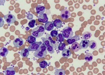
In a new comparative study, researchers traced how this small bile-storage sac and its connecting ducts mature after hatching in three familiar birds: domestic chickens, ducks and pigeons. The work, published in Frontiers in Veterinary Science, documents for the first time a systematic post-hatching timeline of gallbladder histogenesis in chickens and ducks, and pairs it with the alternative biliary plumbing of pigeons, which naturally lack a gallbladder altogether.
The team examined forty clinically healthy birds of both sexes, sixteen chickens, sixteen ducks and eight pigeons, obtained from commercial poultry farms in Menoufia Governorate, Egypt. The birds were divided into four post-hatching age groups: one day, one month, four months and eight months after hatching, with four chickens, four ducks and two pigeons per group. All procedures were approved by the Scientific Research Ethics and Animal Care Committee of the Faculty of Veterinary Medicine at Menoufia University, and birds were euthanized by cervical dislocation following light ether anesthesia in accordance with AVMA guidelines. Post-mortem examination confirmed the absence of gross lesions or biliary disease before any tissue was collected.
The gross anatomy alone delivered a striking contrast. In every chicken and every duck examined, the gallbladder was present, but its shape differed sharply between the species. In chickens it was elongated and tubular, its apex directed caudally, sitting in the cranial half of the visceral surface of the right liver lobe and taking on a dark green appearance from its bile content. In ducks it was ovoid with a cranially directed apex, nestled in a deep cystic fossa on the visceral surface of the right liver lobe without extending to the ventral hepatic border. The pigeon, as expected, showed no gallbladder and no cystic fossa at all.
The ductal architecture revealed three extrahepatic bile ducts in the gallbladder-bearing species: a hepato-cystic duct connecting the right hepatic duct to the gallbladder, a cystico-enteric duct arising from the neck of the gallbladder, and a hepato-enteric duct arising from the right liver lobe. In both chickens and ducks, the cystico-enteric and hepato-enteric ducts, along with two pancreatic ducts, opened onto a common papilla in the distal part of the ascending duodenum. Pigeons, lacking a gallbladder, had no cystico-enteric duct. Instead, their biliary drainage was accomplished by two hepato-enteric ducts: the right duct drained the right liver lobe and opened into the middle portion of the ascending duodenum, while the left duct drained the left liver lobe and opened into the proximal descending duodenum. A single pancreatic duct opened into the ascending duodenum distal to the right hepato-enteric duct opening.
To capture how these structures mature, the researchers fixed gallbladder samples within five minutes of death in ten percent neutral buffered formalin, embedded them in paraffin, and cut serial sections five micrometers thick. The sections were stained with Harris’s hematoxylin and eosin for general histology, Masson’s trichrome for collagen and smooth muscle, Weigert’s elastic stain for elastic fibers, Gomori’s reticulin for reticular fibers, and periodic acid-Schiff combined with Alcian blue at pH 2.5 to detect neutral and acidic mucins simultaneously. Morphometric measurements of epithelial height, villous length and muscularis thickness were taken at ten random fields per section using ImageJ software, and differences among age groups were analyzed by one-way ANOVA followed by Tukey’s post-hoc test, with p less than 0.05 considered significant.
Under the light microscope, the gallbladder wall in both chickens and ducks was composed of four distinct tunics: mucosa, lamina propria-submucosa, muscularis, and serosa or adventitia. At one day post-hatching, the mucosa in both species formed simple longitudinal villus-like folds. The chicken epithelium consisted of low columnar cells with ovoid, basally located vesicular nuclei, while the duck epithelium was made of tall simple columnar cells with large, oval, basally located vesicular nuclei. The lamina propria-submucosa was a thin layer of loose connective tissue rich in collagen and reticular fibers, and the tunica muscularis was a single thin layer of circularly arranged smooth muscle fibers. Even at this earliest stage, the apical cytoplasm of epithelial cells in both species showed intense PAS and Alcian blue positivity, indicating constitutive secretion of mixed neutral and acidic mucins from the very first day of life.
By one month, mucosal complexity had increased in both species. Chicken folds became thicker and branched, lined by simple columnar epithelium with eosinophilic cytoplasm, while duck primary folds became more prominent and exhibited secondary branching, with the tall columnar epithelium displaying extensive apical cytoplasmic vacuolation suggestive of active secretory or absorptive function. Simple tubular gland-like invaginations of the epithelium into the underlying lamina propria-submucosa appeared in both species at this stage. The most dramatic species divergence emerged at four months. In chickens, the mucosa became thinner with longer, branched villus-like folds, and the tunica muscularis differentiated into two distinct layers: a thick inner circular layer and a thinner outer longitudinal layer of smooth muscle. The chicken lamina propria-submucosa also contained numerous lymphoid nodules and compound tubuloalveolar glands, structures entirely absent in ducks, whose muscularis remained a single thick circular layer and whose lymphoid nodules were sparse.
At eight months, the gallbladder reached full histological maturity in both species. The wall was at its maximum thickness, the tunica muscularis was significantly thicker than at all previous stages, and the serosa was the thickest layer, composed of loose connective tissue containing arteries, veins, nerves and adipocytes. Chicken mucosa displayed long, complex, branched villus-like folds, whereas duck folds were comparatively short. Submucosal glands remained prominent in chickens but were never observed in ducks, and the lymphoid nodules seen in chickens at four months had disappeared by eight months, leaving only diffuse lymphatic tissue in the lamina propria. Quantitatively, chicken villus length reached 315.3 plus or minus 19.7 micrometers at eight months, significantly longer than the duck’s 234.4 plus or minus 16.9 micrometers, while epithelial height rose from 12.4 micrometers in day-old chicks to 28.9 micrometers in eight-month-old chickens, and from 14.5 to 35.8 micrometers in ducks. Muscularis thickness climbed from roughly 83 to 98 micrometers in chickens and from about 85 to 120 micrometers in ducks over the same period.
The histochemical results were equally telling. The lining epithelium showed strong PAS and Alcian blue positivity at every examined stage in both species, pointing to persistent mucin production and a constitutive mucosal protective barrier. In chickens, both the surface epithelium and the submucosal glands maintained intense staining through eight months, while duck epithelium shifted to moderate reactivity at four and eight months. The authors interpret this continuous mucin layer as a defense against bile salt cytotoxicity and bacterial translocation, particularly important in precocial birds exposed to environmental pathogens immediately after hatching. They also note that the shift to moderate mucin reactivity in ducks at four months, coinciding with increased bile volume, may reflect a transition from mucus-dependent bile concentration to volume-dependent storage, though they caution that this observation requires further investigation.
The study’s authors frame these findings as a baseline for interpreting biliary pathology in avian models and as a reference for future work on avian hepatobiliary health and disease. They acknowledge the limitations of relying solely on light microscopy, noting that ultrastructural and immunohistochemical analyses of mucin types are needed to fully characterize the protective barrier, and that functional studies correlating bile composition with histological maturity would clarify the physiological significance of the changes described. The gallbladder-to-liver weight ratio declined significantly after one month in both species, indicating allometric growth of the liver relative to the gallbladder, and the disappearance of lymphoid nodules in mature chickens suggests that gallbladder-associated lymphoid tissue may function primarily during juvenile stages. For a structure so often overlooked, the gallbladder turns out to be a remarkably dynamic organ, one whose post-hatching development differs between two domesticated birds in ways that could shape how veterinarians and researchers interpret biliary disease across avian species.
Subject of Research: Post-hatching histogenesis of the gallbladder and extrahepatic bile ducts in chickens, ducks and pigeons
Article Title: Comparative post-hatching histogenesis of gallbladder and extrahepatic bile ducts in chickens, ducks, and pigeons
Article References: Abdel-Megeid, N. S., Fericean, L., Mahmoud, S. F., Elweza, A., Ioan, B.-D., Ostan, M., Rada, O., Magdy, A., & Abdo, M. (2026). Comparative post-hatching histogenesis of gallbladder and extrahepatic bile ducts in chickens, ducks, and pigeons. Frontiers in Veterinary Science, 13, Article 1969438. https://doi.org/10.3389/fvets.2026.1969438
Image Credits: AI Generated
DOI: 10.3389/fvets.2026.1969438
Keywords: gallbladder, histogenesis, chicken, duck, pigeon, bile ducts, avian anatomy, mucin secretion, post-hatching development, comparative histology, veterinary science, biliary system
News Source: Drew Townsend. (November 5, 2026). Chickens and Ducks Build Their Gallbladders Differently After Hatching, While Pigeons Manage Without One. Scienmag.