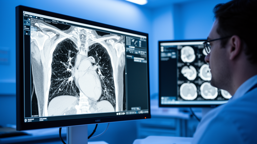
Every year, millions of people undergo chest computed tomography (CT) scans for reasons that have nothing to do with their heart: a persistent cough, suspected pneumonia, cancer screening, or trauma evaluation. These scans capture extraordinarily detailed pictures of the chest, yet most of the information they contain is never analyzed. Radiologists read them for the specific clinical question at hand, and the rest is treated as background noise. Now, a research team led by Yanbo Liang, Kaiyi Shi, and Xiaoqing Yuan at Beijing Friendship Hospital, Capital Medical University, has shown that this discarded data could hold a hidden warning about one of the world’s deadliest diseases: coronary atherosclerosis, the silent buildup of plaque in the arteries that supply the heart muscle itself.
The concept driving the new study, published in BMC Medical Imaging, is known as opportunistic screening. Rather than ordering dedicated tests to look for disease, opportunistic screening extracts medically useful information from imaging exams that were performed for other purposes. Non-contrast chest CT scans are a prime target for this approach because they are already abundant, already paid for, and already expose patients to radiation for a legitimate reason. The researchers asked a deceptively simple question: could the body composition information embedded in these routine scans, when combined with basic clinical data, reliably flag which middle-aged and older adults are carrying coronary atherosclerosis without anyone having ordered a cardiac workup?
To answer it, the team assembled a retrospective cohort of 1,196 adults who had undergone non-contrast chest CT at their institution. The cohort was divided into a training set of 837 patients, used to build the predictive models, and an internal validation set of 359 patients, used to test them. A separate external cohort of 53 patients from another hospital served as an additional, independent check on the models’ generalizability. The study focused specifically on middle-aged and older adults, the demographic where coronary atherosclerosis is most prevalent and where opportunistic detection could have the greatest preventive impact.
The technical heart of the study lies in what the researchers call body composition phenotypes. Using automated image analysis, the team extracted quantitative measurements from the chest CT scans that describe the composition of the patient’s body: the amount of skeletal muscle, its mean attenuation measured in Hounsfield units, the distribution of fat, and other tissue characteristics visible in the chest region. These features were then filtered through a statistical technique called least absolute shrinkage and selection operator (LASSO) regression, which shrinks the coefficients of less informative features toward zero and effectively selects only the most predictive ones. This step is crucial in medical machine learning because it prevents overfitting, where a model memorizes quirks of the training data rather than learning genuine biological signals.
In parallel, the researchers collected clinical variables for each patient, including demographics, medical history, and laboratory parameters such as albumin, liver enzymes, cholesterol fractions, and blood counts, all measured before imaging. These clinical features were screened using logistic regression to identify which ones carried independent predictive value. The team then built four different types of predictive models using the selected features: a generalized linear model (GLM), a support vector machine (SVM), a random forest (RF), and extreme gradient boosting (XGBoost). Each model was tested in three configurations: using clinical data alone, using imaging-derived body composition data alone, and using both combined.
The results were striking. Among all configurations, the combined GLM model, which merged CT-derived body composition phenotypes with clinical information, achieved the best performance. It reached an area under the receiver operating characteristic curve (AUC) of 0.817 (95% confidence interval, 0.788–0.845) in the internal training set, 0.810 (95% CI, 0.760–0.852) in the internal validation set, and 0.726 (95% CI, 0.562–0.851) in the external validation set. An AUC of 0.8 means the model can distinguish between patients with and without coronary atherosclerosis roughly 80 percent of the time, a level of discrimination that approaches the performance of dedicated cardiac risk assessments. The fact that the external validation AUC, while lower, still exceeded 0.7 suggests the model retains meaningful predictive power even when applied to patients from a completely different hospital.
Perhaps the most intriguing finding came from the explainability analysis. The team used SHapley Additive exPlanations (SHAP), a game-theoretic technique that quantifies how much each individual feature contributes to a model’s prediction for each patient. SHAP analysis revealed that skeletal muscle mean attenuation, a measure of muscle quality rather than quantity, was the single strongest contributor in the image-based model. This suggests that the health of a patient’s muscle tissue, as reflected in how bright or dark it appears on a CT scan, carries a surprisingly strong signal about the state of their coronary arteries. Low muscle attenuation is associated with fatty infiltration and poor muscle quality, which in turn correlate with metabolic dysfunction, inflammation, and vascular disease.
The clinical implications of this work are substantial. Coronary atherosclerosis is a leading cause of death worldwide, yet it often progresses silently for decades before producing symptoms. Current screening approaches, such as coronary artery calcium scoring, require dedicated CT protocols with specific gating and radiation exposure. Opportunistic screening, by contrast, requires no additional scans, no additional radiation, and no additional cost beyond the computational analysis itself. If validated in larger prospective studies, the approach could allow radiologists to append an automated cardiovascular risk flag to routine chest CT reports, prompting clinicians to order further cardiac evaluation for patients who might otherwise go undetected.
The study does have limitations that temper enthusiasm. The retrospective design means the models were built and tested on existing data rather than prospectively assigned patients, and the external validation cohort was small at only 53 patients. The AUC drop from internal to external validation, while expected, highlights the challenge of generalizing machine learning models across different hospitals with different patient populations and scanning protocols. The researchers also note that non-contrast chest CT scans contain a large amount of background redundant information, and their clinical utilization is currently insufficient, which is precisely the gap this study aims to close.
Nevertheless, the work represents a compelling step toward a future where every medical image is mined for all the information it contains, not just the answer to the question that prompted it. Features derived from non-contrast chest CT, combined with clinical information, can opportunistically identify coronary atherosclerosis risk in middle-aged and older adults, extending the utility of chest CT beyond its primary diagnostic purpose. As the researchers conclude, this approach could transform the humble chest CT from a single-purpose diagnostic tool into a multipurpose screening instrument, quietly watching for hidden disease every time a patient steps into the scanner for something else entirely.
Subject of Research: Opportunistic screening chest CT-derived body composition phenotypes combined with clinical information to identify coronary atherosclerosis risk in middle-aged and older adults.
Article Title: Identification of the risk of coronary atherosclerosis using opportunistic screening chest CT-derived body composition phenotypes combined with clinical information in middle-aged and older adults
Article References: Liang, Y., Shi, K., Yuan, X., Yang, H., Wang, Z., Jing, C., Sun, R., Qu, J., Qin, J., & Lv, H. (2026). Identification of the risk of coronary atherosclerosis using opportunistic screening chest CT-derived body composition phenotypes combined with clinical information in middle-aged and older adults. BMC Medical Imaging. https://doi.org/10.1186/s12880-026-02718-0
Image Credits: AI Generated
DOI: 10.1186/s12880-026-02718-0
Keywords: chest CT, coronary atherosclerosis, body composition, machine learning, opportunistic screening, medical imaging, heart disease risk, Identification, risk, coronary, atherosclerosis, opportunistic
News Source: Ophelia Keating. (October 17, 2026). Chest CT Scans Could Secretly Reveal Your Heart Attack Risk Years Early. Scienmag.