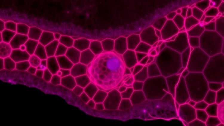
Every cell in a developing tissue faces a decision. In the embryonic skin of the African clawed frog, Xenopus laevis, multipotent progenitors must choose among becoming multiciliated cells, goblet cells, ionocytes, small secretory cells, or basal stem cells. Biologists have long mapped these choices through gene expression, using single-cell RNA sequencing to read out the molecular programs that define each fate. But a cell is more than its transcriptome. It also has a shape, a position, and a pattern of movement, and a new study published in Molecular Systems Biology shows that these morphodynamic features, tracked continuously over many hours of live imaging, carry enough information to predict which fate a cell will ultimately adopt.
The research, led by Mari Tolonen, Ziwei Xu, and Jakub Sedzinski of the University of Copenhagen together with Bianca Dumitrascu’s group at Columbia University, tackled a problem that has long frustrated developmental biologists: how to quantify the behavior of thousands of individual cells inside a densely packed, actively remodeling tissue. Morphology is often treated as a downstream consequence of fate decisions, a passive readout of the genetic program. The team set out to test a bolder idea, namely that the dynamic geometry of a cell might serve as a complementary, and potentially predictive, indicator of its state, in the same way that transcriptomic profiles define molecular identity.
Their experimental system was the Xenopus mucociliary epithelium, a bilayered embryonic skin that closely resembles the cellular composition of the mammalian airway. The researchers dissected animal cap explants from embryos at early blastula stages, when the tissue was still multipotent, and mounted them on fibronectin-coated dishes where they flattened into stratified epithelia amenable to long-term optical access. Embryos had been injected with messenger RNAs encoding membrane-localized mNeonGreen and nuclear H2B-RFP, allowing both cell boundaries and nuclei to be visualized. Volumetric confocal time-lapse imaging then captured the full course of differentiation, from NF stage 9 through stage 30, with z-stacks acquired every three to five minutes over 16 to 22 hours across three independent experiments.
Turning those raw movies into quantitative data required a substantial computational pipeline. Nuclei were segmented in three dimensions using a custom-trained StarDist model, while membrane signals were first denoised with a CARE network and then segmented slice by slice with Cellpose, with individual 2D contours stitched into 3D volumes based on maximal pairwise overlap. Nuclear objects were tracked over time using TrackMate, and the team integrated a convolutional neural network called Oneat to detect cell division events, which were used to impose branching points on trajectories. This combination substantially improved mitosis detection compared with TrackMate’s native algorithm, raising branching correctness from 0.122 to 0.328. On average, trajectories spanned at least 9.5 hours in a third of cells, providing sufficient temporal coverage for the analyses that followed.
From the segmentations and tracks, the researchers computed a rich set of time-resolved features for each cell: three-dimensional shape descriptors derived from point clouds resampled from surface meshes, including surface area, eccentricity, and orientation; conventional two-dimensional descriptors such as area, Feret diameter, and solidity taken from each membrane object’s central slice; nuclear shape metrics; spatial position; and instantaneous movement. The result was a high-dimensional morphodynamic feature space, conceptually analogous to a transcriptomic state vector, in which each cell’s identity at each moment was represented by dozens of quantitative measurements.
When the team explored this feature space with unsupervised methods, the picture was sobering. Principal component analysis of more than 4,200 cell trajectories revealed no distinct clusters corresponding to cell types or developmental stages. Cells were distributed continuously, with only slight shifts over time, and standard clustering metrics such as the Adjusted Rand Index and Normalized Mutual Information were uniformly low. A simple K-nearest-neighbor classifier reached a mean accuracy of just 65.8 percent against a stratified baseline of 54.0 percent, with balanced accuracy of only 28.1 percent. Unlike cultured cells, where shape heterogeneity is high and morphology alone can resolve distinct states, cells in this densely packed epithelium share constrained shapes and largely collective movement, making them hard to distinguish by any single feature.
The breakthrough came from supervised learning combined with lineage backtracking. After live imaging ended, the samples were fixed and immunostained for markers of multiciliated cells, goblet cells, small secretory cells, and basal cells, while ionocytes were identified by their characteristic morphology and lack of marker expression. By aligning the immunostained images with the final live-imaging frame, the team assigned terminal fates to roughly 75 percent of all tracked trajectories, producing a ground-truth dataset of 2,576 curated lineages. They then trained an XGBoost gradient-boosted classifier and a multinomial logistic regression model, treating each cell at each time point as an independent observation labeled by its track’s endpoint fate, with absolute experimental time included as an additional input.
The supervised models achieved moderate but consistent performance across 20 independent train-test splits. XGBoost reached a mean accuracy of 80.6 percent and a balanced accuracy of 61.4 percent, while logistic regression achieved 70.3 percent accuracy with a comparable balanced accuracy of 62.9 percent. Performance was strongest for the abundant epithelial lineages: basal cells scored an F1 of 0.90, goblet cells 0.72, and multiciliated cells 0.56 under XGBoost, whereas the rare ionocytes and small secretory cells remained difficult to classify despite class balancing with SMOTE-based oversampling and undersampling. Notably, classifier confidence and accuracy rose steadily during development, with mean confidence increasing from 0.80 at around five hours to 0.89 by 22 hours, and class-balanced accuracy climbing from 50.4 to 75.1 percent. Intriguingly, predictions for multiciliated cells peaked at roughly 15 hours, with 80.6 percent accuracy, and then declined, suggesting these cells reach a morphodynamic plateau before terminal maturation, after which their features increasingly overlap with those of goblet cells.
Feature importance analysis revealed which aspects of cellular behavior carried the predictive signal. Normalized Z position, reflecting a cell’s height within the apico-basal axis of the epithelium, was the single most informative feature, consistent with the invariant layer preferences of different lineages: goblet cells remain in the superficial layer, basal stem cells stay confined to the base, and intercalating lineages ascend. The nucleus-to-membrane centroid offset was the most predictive morphological variable in the XGBoost model, likely capturing the transition from a flat, migratory configuration to a vertically oriented shape during radial intercalation. Absolute experimental time was also a strong predictor, particularly for small secretory cells, which emerge late in development. Perhaps most strikingly, movement features contributed almost nothing to fate prediction, apparently because migration within the tissue is largely collective rather than fate-specific. Nuclear shape features proved more informative than membrane shape, which the authors attribute to the higher segmentation fidelity of nuclei.
The broader significance of this work lies in what it suggests about the nature of cell state itself. Fate, the study indicates, is not defined by discrete morphological states that cells snap into; instead, morphodynamic signatures of identity emerge gradually along continuous, overlapping trajectories, and only become legible through time-integrated, supervised analysis. Because morphodynamic tracking provides dense temporal sampling with positional context, something neither single-cell RNA sequencing nor spatial transcriptomics can fully deliver, it could complement molecular fate mapping by filling temporal and spatial gaps. The authors note that their pipeline, built from open tools including StarDist, Cellpose, TrackMate, and Oneat, can be extended to other developing tissues and combined with transcriptomics or targeted perturbations to uncover causal links between gene expression, signaling, and phenotype. Future models that incorporate neighboring cells and local tissue geometry may sharpen predictions further. For now, the message is clear: the shape a cell takes, where it sits, and when it does so are not mere consequences of its destiny but readable, quantitative clues to what that destiny will be.
Subject of Research: Machine learning prediction of cell fate from single-cell morphodynamic features during Xenopus mucociliary epithelial differentiation
Article Title: Single-cell morphodynamics predict cell fate decisions during mucociliary epithelial differentiation
Article References: Tolonen, M., Xu, Z., Beker, O., Kapoor, V., Dumitrascu, B., & Sedzinski, J. (2026). Single-cell morphodynamics predict cell fate decisions during mucociliary epithelial differentiation. Molecular Systems Biology, 22(7), 1040-1069. https://doi.org/10.1038/s44320-026-00212-x
Image Credits: AI Generated
DOI: 10.1038/s44320-026-00212-x
Keywords: cell fate, cell differentiation, Xenopus laevis, mucociliary epithelium, live imaging, morphodynamics, machine learning, XGBoost, cell tracking, segmentation, radial intercalation, developmental biology
Cite Scienmag News
APA
MLA
Chicago
Drew Townsend. (October 1, 2026). Cell Shape and Position Predict Fate in a Developing Epithelium. Scienmag. https://scienmag.com/cell-shape-and-position-predict-fate-in-a-developing-epithelium/
Drew Townsend. “Cell Shape and Position Predict Fate in a Developing Epithelium.” Scienmag, 1 October 2026, https://scienmag.com/cell-shape-and-position-predict-fate-in-a-developing-epithelium/. Accessed 1 October 2026.
Drew Townsend. “Cell Shape and Position Predict Fate in a Developing Epithelium.” Scienmag. October 1, 2026. https://scienmag.com/cell-shape-and-position-predict-fate-in-a-developing-epithelium/
Copy citation
Download RIS
Tags: cell behavior quantificationcell differentiationcell fatecell fate predictioncell morphodynamicscell patterningcell shape and position in epitheliumcell trackingdevelopmental biologylive cell imaginglive imagingMachine learningmorphodynamicsmorphogenetic pattern analysismucociliary epitheliummultipotent progenitorsradial intercalationsegmentationSingle-Cell RNA Sequencingtissue remodelingXenopus laevisXenopus laevis embryonic skinXGBoost