• HOME
  • NEWS
  • EXPLORE
    • CAREER
      • Companies
      • Jobs
    • EVENTS
    • iGEM
      • News
      • Team
    • PHOTOS
    • VIDEO
    • WIKI
  • BLOG
  • COMMUNITY
    • FACEBOOK
    • INSTAGRAM
    • TWITTER
Monday, September 22, 2025
BIOENGINEER.ORG
No Result
View All Result
  • Login
  • HOME
  • NEWS
  • EXPLORE
    • CAREER
      • Companies
      • Jobs
        • Lecturer
        • PhD Studentship
        • Postdoc
        • Research Assistant
    • EVENTS
    • iGEM
      • News
      • Team
    • PHOTOS
    • VIDEO
    • WIKI
  • BLOG
  • COMMUNITY
    • FACEBOOK
    • INSTAGRAM
    • TWITTER
No Result
View All Result
Bioengineer.org
No Result
View All Result
Home NEWS Science News Chemistry

Cell membranes in super resolution

Bioengineer by Bioengineer
December 2, 2020
in Chemistry
Reading Time: 2 mins read
0
IMAGE
Share on FacebookShare on TwitterShare on LinkedinShare on RedditShare on Telegram

IMAGE

Credit: (Picture: Sauer group / University of Würzburg)

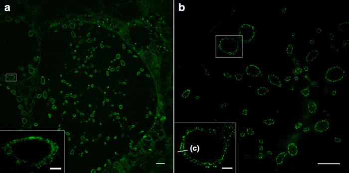
Expansion microscopy (ExM) enables the imaging of cells and their components with a spatial resolution far below 200 nanometres. For this purpose, the proteins of the sample under investigation are cross-linked into a swellable polymer. Once the interactions between the molecules have been destroyed, the samples can be expanded many times over with water. This allows detailed insights into their structures.

“This method was previously limited to proteins. In the journal Nature Communications we are now presenting a way of expanding lipids and thus cell membranes,” says Professor Markus Sauer, an expert in super-resolution microscopy from the Biocentre of Julius-Maximilians-Universität (JMU) Würzburg in Bavaria, Germany. JMU professors Thomas Rudel (microbiology) and Jürgen Seibel (chemistry) are also involved in the publication.

Synthetic lipids are marked and expanded

Jürgen Seibel’s team has synthesised functionalised sphingolipids, which are an important component of cell membranes. If these lipids are added to cell cultures, they are incorporated into the cell membranes. They can then be marked with a dye and expanded four to ten times in a swellable polymer.

The JMU researchers show that this method – in combination with structured illumination microscopy (SIM) – makes it possible for the first time to image different membranes and their interactions with proteins with a resolution of 10 to 20 nanometres: cell membranes, the outer and inner cell nuclear membrane and also the membranes of intracellular organelles such as mitochondria.

Focusing on bacteria and viruses

The sphingolipids also integrate highly efficiently into the membranes of bacteria. This means that, for the first time, pathogens such as Neisseria gonorrhoeae, Chlamydia trachomatis and Simkania negevensis can now be visualised in infected cells with a resolution that was previously only achieved using electron microscopy. Even the inner and outer membranes of Gram-negative bacteria can be distinguished from each other.

“With the new super-resolution microscopic methods, we now want to investigate bacterial infection mechanisms and causes of antibiotic resistance. What we learn in the process could possibly be used for improved therapies,” says Professor Thomas Rudel, an expert on bacterial infections.

The sphingolipids might also integrate into the membrane of viruses. If this is successful, the interactions of corona viruses with cells could be studied for the first time with high resolution light microscopy.

###

Media Contact
Dr. Markus Sauer
[email protected]

Original Source

https://go.uniwue.de/exm-membranes

Related Journal Article

http://dx.doi.org/10.1038/s41467-020-19897-1

Tags: BacteriologyBiologyBiomechanics/BiophysicsCell BiologyInfectious/Emerging DiseasesMicrobiologyMolecular BiologyVirology
Share12Tweet8Share2ShareShareShare2

Related Posts

New Study Warns Seasonal Freeze–Thaw Cycles Could Cause “Green” Biochar to Release Toxic Metals

New Study Warns Seasonal Freeze–Thaw Cycles Could Cause “Green” Biochar to Release Toxic Metals

September 20, 2025
blank

Gravitino Emerges as a Promising New Candidate for Dark Matter

September 19, 2025

Advancing Quantum Chemistry: Enhancing Accuracy in Key Simulation Methods

September 19, 2025

Neutrino Mixing in Colliding Neutron Stars Alters Merger Dynamics

September 19, 2025
Please login to join discussion

POPULAR NEWS

  • blank

    Breakthrough in Computer Hardware Advances Solves Complex Optimization Challenges

    156 shares
    Share 62 Tweet 39
  • Physicists Develop Visible Time Crystal for the First Time

    68 shares
    Share 27 Tweet 17
  • Tailored Gene-Editing Technology Emerges as a Promising Treatment for Fatal Pediatric Diseases

    49 shares
    Share 20 Tweet 12
  • Scientists Achieve Ambient-Temperature Light-Induced Heterolytic Hydrogen Dissociation

    48 shares
    Share 19 Tweet 12

About

We bring you the latest biotechnology news from best research centers and universities around the world. Check our website.

Follow us

Recent News

Key Drivers of Corporate Governance in Burundi’s Cooperatives

Revolutionizing Sustainable Construction: The Role of Cardboard and Earth

TMolNet: Revolutionizing Molecular Property Prediction

  • Contact Us

Bioengineer.org © Copyright 2023 All Rights Reserved.

Welcome Back!

Login to your account below

Forgotten Password?

Retrieve your password

Please enter your username or email address to reset your password.

Log In
No Result
View All Result
  • Homepages
    • Home Page 1
    • Home Page 2
  • News
  • National
  • Business
  • Health
  • Lifestyle
  • Science

Bioengineer.org © Copyright 2023 All Rights Reserved.