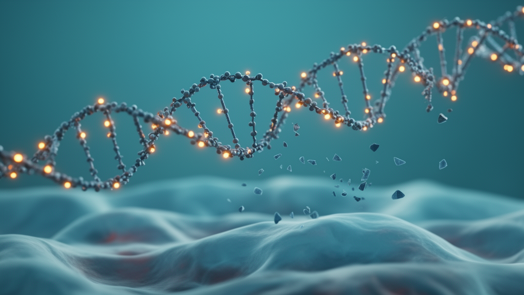
A single tube of blood contains far more than cells and proteins. It also carries cell-free DNA (cfDNA), short fragments of genetic material shed into the bloodstream as cells die. For more than a decade, researchers have mined this circulating DNA for cancer clues, reading its chemical marks and its physical shattering patterns as two separate languages. Now a study published in Genome Medicine has shown that these two languages are deeply intertwined, and that reading them together, in a biologically grounded way, can produce cancer diagnostic models that are both powerful and interpretable.
The research, led by Yunze Wang, Yuying Hou, Xinyu Liu, Wen Zhang, and Xionghui Zhou of Huazhong Agricultural University together with Qingjiao Li of Sun Yat-Sen University’s Eighth Affiliated Hospital, tackles a practical problem that has quietly limited the field. Whole-genome methylation sequencing of plasma cfDNA is attractive because a single assay can capture both DNA methylation and fragment-level features. But nobody had rigorously tested whether the chemical steps used to read methylation actually preserve the fragmentomic signals that other methods rely on.
The team systematically compared two methylation library preparation protocols, whole-genome bisulfite sequencing (WGBS) and enzymatic methyl sequencing (EM-seq), against conventional whole-genome sequencing (WGS). Their verdict was nuanced. Canonical cfDNA fragmentomic patterns, such as characteristic fragment size distributions and end motifs, were partially preserved across the methylation protocols, but the degree of preservation varied from feature to feature. In other words, methylation sequencing is not a perfect stand-in for WGS when it comes to fragmentation, and researchers combining both modalities from one assay need to know exactly which signals survive the chemistry.
Beyond the technical audit, the study probed the biology linking the two modalities. DNA methylation levels turned out to be associated with characteristic fragment length and end-motif patterns, confirming that how a DNA molecule is chemically decorated influences how it is cut, protected, and degraded in the blood. This coupling is not a nuisance; it is information. Tumor cells, with their altered chromatin and epigenetic landscapes, release DNA that carries coordinated epigenetic and physical fingerprints, and the study set out to organize those fingerprints into a framework a computer model can actually use.
The organizing principle the researchers chose is the methylation haplotype block, or MHB. Rather than treating each CpG site or each fragment independently, the team delineated MHBs directly from plasma cfDNA. These blocks, stretches of the genome where methylation states tend to be inherited together across reads, exhibited tissue-associated regulatory features and distinct patterns of chromatin accessibility, repeat enrichment, and methylation heterogeneity. That makes them biologically interpretable units, anchored in real regulatory architecture rather than arbitrary genomic windows.
Within these MHB regions, methylation and fragmentomic signals showed coordinated yet complementary patterns. Methylation tells the model which tissue the DNA came from; fragmentation tells it how that DNA was processed and cleared. Neither signal alone captures the full picture, but together they form a richer portrait of what is happening in a patient’s body. This insight guided the construction of FAME, the Fragmentomics And Methylation Ensemble, an interpretable multimodal diagnostic model that integrates MHB-centered methylation and fragmentomic features for cancer detection and tissue-of-origin prediction.
The researchers did not stop at a compact model. They extended FAME with complementary genome-wide methylation and fragmentomic features, producing FAME-GW, a genome-wide-augmented version. Across cross-validation, independent validation cohorts, and head-to-head comparisons with established methods including THEMIS (THorough Epigenetic Marker Integration Solution), SPOT-MAS (screening for the presence of tumor by methylation and size), and DELFI (DNA evaluation of fragments for early interception), the FAME-based models achieved robust diagnostic performance. FAME delivered a compact and interpretable representation, while FAME-GW delivered the strongest overall predictive performance.
What makes this work stand out in the crowded liquid biopsy field is its insistence on interpretability. Many high-performing cancer detection models are black boxes, blending thousands of features in ways clinicians cannot inspect. By centering the analysis on MHBs, units with known regulatory and chromatin properties, the FAME framework lets researchers trace a prediction back to biologically meaningful regions. A clinician or scientist can ask which blocks drove a cancer call and what their methylation and fragmentation patterns look like, rather than trusting an opaque score.
The study also carries a practical message for anyone designing cfDNA assays. Because methylation library preparation partially preserves fragmentomic signals, a single whole-genome methylation sequencing run can serve double duty, capturing both epigenetic and fragment-level information from one precious plasma sample. That is a meaningful efficiency gain for multi-cancer early detection programs, where sample volume is limited and every read counts. But the feature-by-feature variation in preservation means assay developers must validate each fragmentomic signal they intend to use under their specific protocol.
The work, published on 15 October 2026 as an open-access article in Genome Medicine, was supported by the Fundamental Research Funds for the Central Universities and the Huazhong Agricultural University Scientific and Technological Self-innovation Foundation. The in-house cohort drew healthy controls from The Eighth Affiliated Hospital of Sun Yat-sen University and gastric cancer samples from Fujian Medical University Union Hospital, with ethics approval and written informed consent obtained for all participants. As methylation haplotype blocks move from a research framework toward clinical assay design, the study’s central conclusion is likely to shape the next generation of cfDNA diagnostics: the epigenetic and fragmentomic stories written in circulating DNA are not separate chapters but a single, coupled narrative, and models that read them together, in interpretable units, are the ones most likely to reach the clinic.
Subject of Research: Integrating coupled methylation and fragmentomic signals in plasma cell-free DNA for interpretable cancer diagnosis
Article Title: Decoding coupled epigenetic-fragmentomic signals in circulating cell-free DNA for interpretable cancer diagnosis
Article References: Wang, Y., Hou, Y., Liu, X., Li, Q., Zhang, W., & Zhou, X. (2026). Decoding coupled epigenetic-fragmentomic signals in circulating cell-free DNA for interpretable cancer diagnosis. Genome Medicine, 18(1), Article 154. https://doi.org/10.1186/s13073-026-01793-w
Image Credits: AI Generated
DOI: 10.1186/s13073-026-01793-w
Keywords: cell-free DNA, DNA methylation, fragmentomics, methylation haplotype blocks, liquid biopsy, cancer detection, whole-genome bisulfite sequencing, EM-seq, FAME model, tissue of origin, Genome Medicine, interpretable machine learning
News Source: Nathaniel Bowman. (October 16, 2026). Cell-Free DNA Study Reveals How Methylation and Fragmentation Team Up for Cancer Detection. Scienmag.