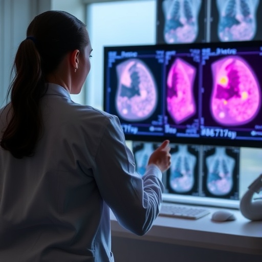
In a groundbreaking advancement at the intersection of artificial intelligence and medical imaging, researchers from The City College of New York (CCNY) together with collaborators from Memorial Sloan Kettering Cancer Center (MSKCC) have unveiled an innovative AI model designed to detect breast cancer in MRI scans with exceptional accuracy. This new deep learning system not only identifies the presence of malignancies but also accurately localizes tumors within the breast tissue—a critical factor in diagnosis and treatment planning. Detailed in the prestigious journal Radiology: Artificial Intelligence, this development signals a pivotal shift toward more interpretable, transparent, and widely accessible AI tools in oncology.
Breast cancer detection has historically relied heavily on mammography as the frontline screening method due to its widespread availability, cost-effectiveness, and reasonable sensitivity levels. However, mammography’s limitations, especially in patients with dense breast tissue, have paved the way for Magnetic Resonance Imaging (MRI) to emerge as a more sensitive alternative. Despite MRI’s superior sensitivity, variable imaging protocols and limited dataset sizes have posed significant challenges for developing robust AI models tailored to this modality. The CCNY-MSKCC team tackled these hurdles head-on by curating the largest breast MRI dataset to date, ensuring their AI system generalizes well across heterogeneous clinical environments.
One of the most striking features of this model lies in its interpretability and openness. Unlike many current deep learning approaches, which tend to operate as ‘black boxes’ with limited transparency, this algorithm was designed to provide visual and clinical interpretability, highlighting suspected tumor regions clearly within MRI scans. In addition, the model has been publicly released, addressing a major pain point in AI research—restricted access and lack of external validation—thus empowering independent researchers and clinicians to evaluate, refine, and build upon this technology. This approach fosters collaborative progress and transparency in a field often criticized for proprietary constraints.
.adsslot_uoqbMJweEF{ width:728px !important; height:90px !important; }
@media (max-width:1199px) { .adsslot_uoqbMJweEF{ width:468px !important; height:60px !important; } }
@media (max-width:767px) { .adsslot_uoqbMJweEF{ width:320px !important; height:50px !important; } }
ADVERTISEMENT
Technically, the model utilizes convolutional neural networks (CNNs) trained on a meticulously annotated dataset comprising thousands of breast MRI images sourced from two distinct clinical institutions. This multi-center data approach is particularly important for capturing the variability across different MRI scanners, imaging sequences, and patient demographics, thereby enhancing the model’s robustness. The architectural design of this AI system integrates localization and classification tasks into a unified framework, enabling simultaneous detection of lesions and precise tumor delineation—a crucial capability for targeted treatment modalities such as surgical planning or radiation therapy.
The AI’s success is particularly significant in light of evolving clinical guidelines recommending expanded MRI utilization for breast cancer screening, especially for women categorized as high risk or those with radiologically dense breast tissue. As MRI-based screening programs expand, reliable and accessible automated interpretation tools become essential to ensure timely, accurate results. This AI model, by seamlessly integrating with existing workflows and offering open-source availability, is well-positioned to support broader adoption of MRI in breast cancer screening paradigms.
Developing such an advanced system required a synergistic collaboration among multidisciplinary experts. The research team at CCNY, led by Professor Lucas C. Parra of Biomedical Engineering, combined expertise in machine learning and medical imaging, while MSKCC’s radiology specialists contributed critical clinical insights and validation. Such cross-institutional partnerships highlight the importance of combining computational innovation with clinical acumen when confronting complex medical challenges like cancer detection.
Beyond cancer detection, the researchers envision this platform as a foundation for future developments in precision oncology. Incorporating machine learning techniques to analyze longitudinal imaging data may enable more accurate risk stratification, monitoring of tumor progression, and evaluation of treatment response. This trajectory aligns with the broader movement toward personalized medicine, where data-driven tools support tailored interventions to improve patient outcomes while reducing unnecessary procedures.
Funding from a substantial $4 million NIH grant supports the ongoing refinement and validation of this AI technology. The project, aptly named “Machine learning for risk-adjusted breast MRI screening,” aims to optimize cancer detection efficacy while minimizing the burden of frequent screenings, especially for women at elevated risk. This objective reflects an awareness of patient-centered care, reducing anxiety and overdiagnosis, and emphasizing timely interventions for those who need them most.
The open-source nature of the model represents a deliberate choice to democratize cutting-edge research tools and accelerate their clinical translation. By making code and training data publicly accessible, the CCNY-MSKCC team encourages transparency and reproducibility, elements often missing in AI studies that hinder real-world deployment. This precedent could signal a new standard in medical AI research, where collaboration and openness drive shared improvements rather than siloed progress.
Looking forward, the integration of this AI system into clinical MRI workflows will require further validation through prospective clinical trials and regulatory reviews. However, the promising results published thus far foreshadow a future where machine learning augments radiologists’ expertise, bringing significant advantages in early cancer detection and localization. In turn, this could translate into more personalized, timely treatments and ultimately improved survival rates for breast cancer patients worldwide.
In sum, this breakthrough represents a leap forward in harnessing artificial intelligence for one of the most pressing public health challenges: early breast cancer detection. By combining state-of-the-art deep learning methodologies with large, diverse clinical datasets and a commitment to open science, the CCNY and MSKCC collaboration has crafted a powerful tool that holds promise for enhancing diagnostic accuracy, clinical efficiency, and patient care in the rapidly evolving field of oncologic imaging.
Subject of Research: Human tissue samples
Article Title: High-performance Open-source AI for Breast Cancer Detection and Localization in MRI
News Publication Date: 25-Jun-2025
Web References:
Memorial Sloan Kettering Cancer Center
Radiology: Artificial Intelligence Journal Article
CCNY Biomedical Engineering
CCNY News – NIH Grant Project
Keywords: Artificial intelligence, breast cancer detection, MRI imaging, deep learning, tumor localization, medical imaging, radiology, open-source AI, biomedical engineering, cancer screening, machine learning, precision oncology
Tags: advancements in breast cancer diagnosisartificial intelligence in radiologybreast cancer detection AICCNY MSKCC collaborationchallenges in breast cancer imagingDeep Learning in Oncologyenhancing MRI sensitivity for cancerinnovations in cancer treatment planninginterpretable AI tools in healthcarelarge datasets for AI trainingMRI tumor localizationopen-source medical imaging technology


