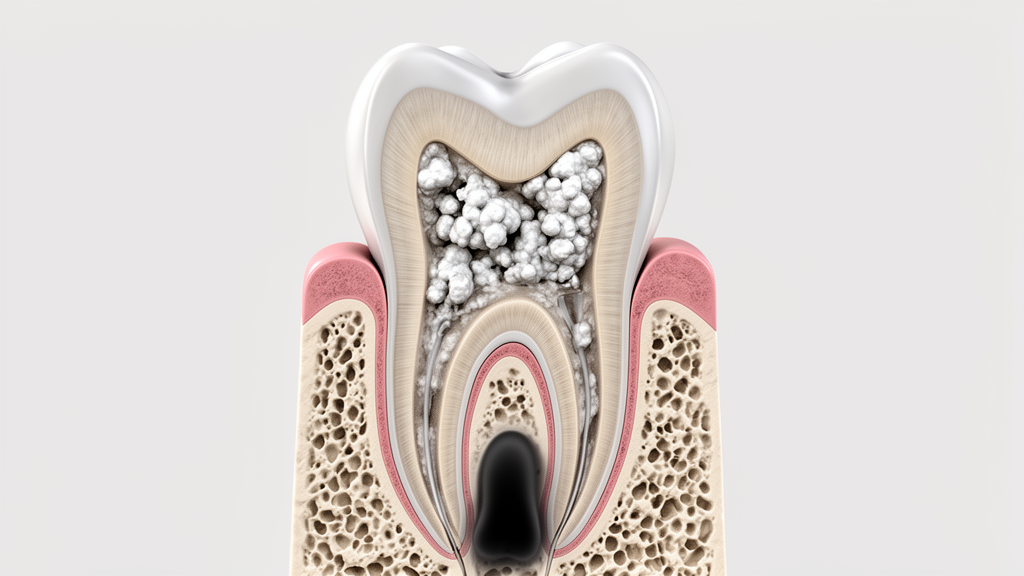
A retrospective dental imaging study has evaluated the radiographic features linked to apical radiolucencies in permanent teeth exhibiting pulpal calcifications. Evaluating three-dimensional cone-beam computed tomography scans, researchers identified apical periodontitis in 18 out of 501 examined teeth, establishing a baseline prevalence of 3.6 percent in the evaluated sample. The primary investigation centered on examining how demographic variables and structural dental modifications associate with inflammatory radiographic presentations at the tooth root apex.
Apical periodontitis represents an inflammatory condition primarily triggered by microbial colonization of the root canal system. Its pathological development can also be influenced by broader structural and periodontal alterations, including pulp canal obliteration, root resorption processes, and alveolar bone reduction. Understanding the precise interplay among these radiographic findings remains critical for diagnostic evaluation, prompting the research team to investigate associations in teeth characterized by pulpal calcifications using high-resolution volumetric imaging.
The methodological framework involved reviewing 203 cone-beam computed tomography examinations obtained from 203 individual patients. Across these scans, 501 permanent teeth presented with pulpal calcifications and were designated as the definitive units of analysis. Each tooth underwent detailed evaluation for radiographically confirmed apical periodontitis, pulp canal obliteration, discrete pulp stones, external root resorption, periodontal ligament widening, and surrounding alveolar bone height.
To analyze the imaging observations, investigators initiated univariable statistical assessments across all measured parameters. These evaluations were subsequently followed by multivariable logistic regression modeling utilizing generalized estimating equations to appropriately account for clustering effects at the individual patient level. Additionally, sensitivity testing was conducted using Firth’s penalized logistic regression specifically for pulp canal obliteration and pulp stones due to statistical data separation.
During initial univariable testing, apical periodontitis showed statistically significant associations with maxillary tooth location, tooth category, periodontal ligament space widening, external root resorption, pulp canal obliteration, and elevated alveolar bone loss, with all comparisons yielding P values below 0.05. These unadjusted associations highlighted an initial cluster of structural and positional variables correlated with periapical pathology in calcified teeth.
Subsequent multivariable generalized estimating equation regression revealed distinct independent associations. Female sex was associated with reduced odds of presenting with apical periodontitis. Concurrently, periodontal ligament space widening and external root resorption remained independently associated with the presence of apical periodontitis after adjusting for confounding variables within the multivariable framework.
Conversely, Firth’s penalized logistic regression yielded imprecise effect estimates accompanied by wide confidence intervals when evaluating pulp canal obliteration and pulp stones. Consequently, the independent association between these specific calcification forms and apical periodontitis remained inconclusive. Furthermore, while periodontal ligament widening and external root resorption demonstrated large statistical effect estimates, both measures exhibited wide confidence intervals.
The authors concluded that among permanent teeth with pulpal calcifications, widening of the periodontal ligament space and external root resorption are independently associated with apical radiolucencies, whereas pulp canal obliteration and pulp stones showed no independent association. Because wide confidence intervals characterized the effect estimates, the researchers emphasized that all identified associations require cautious interpretation.
Subject of Research: Radiographic characteristics associated with apical radiolucencies in teeth with pulpal calcifications
Article Title: Radiographic factors associated with apical radiolucencies in teeth with pulpal calcifications: a retrospective CBCT study
Article References: Madfa, A. A., Al-Enizy, A. S., Alotaibi, Y. A., Alyahya, R. S., Bhagwath, S. S., Alshammari, S. S., & Alshammari, A. F. (2026). Radiographic factors associated with apical radiolucencies in teeth with pulpal calcifications: a retrospective CBCT study. BMC Oral Health. https://doi.org/10.1186/s12903-026-10450-y
Image Credits: AI Generated
DOI: 10.1186/s12903-026-10450-y
Keywords: cone-beam computed tomography, apical periodontitis, pulpal calcification, pulp canal obliteration, pulp stones, external root resorption, periodontal ligament widening, alveolar bone loss, endodontics, dental imaging, generalized estimating equations, retrospective study, oral radiology, periapical radiolucency, tooth calcification
News Source: Ophelia Keating. (November 20, 2026). CBCT Study Evaluates Apical Radiolucencies in Teeth With Pulpal Calcifications. Scienmag.