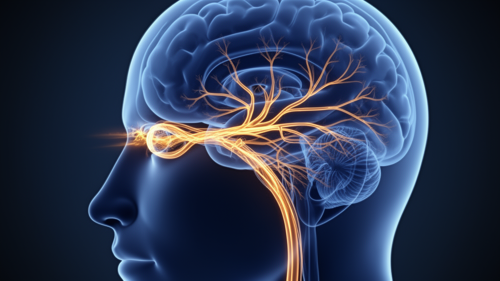
A 65-year-old woman presented with a complex constellation of ocular and neurologic symptoms following a varicella-zoster virus infection, leading to a diagnosis of herpes zoster ophthalmicus complicated by orbital apex syndrome and virus-associated meningitis. The case, detailed in a report published in BMC Ophthalmology, illustrates the rare but severe progression of shingles from the eye to the central nervous system. The patient initially developed pain and a vesicular eruption in the distribution of the left ophthalmic division of the trigeminal nerve, a pattern characteristic of herpes zoster ophthalmicus. As the condition advanced, she exhibited left-sided ptosis, proptosis, esotropia, restricted ocular motility, and decreased vision, indicating significant involvement of the orbital apex.
Upon admission, clinical measurements revealed distinct asymmetries between the patient’s eyes. Visual acuity in the left eye was recorded at 0.16 and did not improve with refraction, suggesting a structural or neurological deficit rather than a simple refractive error. Exophthalmometry measurements showed a protrusion of 20 mm in the left eye compared to 17 mm in the right eye, confirming the proptosis observed clinically. The left pupil was dilated and unreactive to light, a finding that points to parasympathetic nerve involvement within the orbital apex. These physical signs, combined with the history of vesicular rash, prompted a thorough investigation into the extent of the viral spread.
Imaging and laboratory analyses were critical in defining the scope of the infection. Orbital magnetic resonance imaging performed on May 16, 2025, revealed thickening and abnormal signal intensity in the left optic nerve and multiple extraocular muscles.
During her hospitalization, the patient’s condition evolved with the emergence of new neurologic symptoms, including headache and recurrent nausea and vomiting. To investigate this progression, cerebrospinal fluid analysis was conducted.
Advanced molecular techniques were employed to identify the specific viruses involved in the patient’s condition. Probe-capture high-throughput sequencing of the cerebrospinal fluid detected the presence of both varicella-zoster virus and Epstein-Barr virus.
The treatment strategy was multifaceted, targeting both the viral infection and the associated inflammatory response. The patient received systemic antiviral therapy to combat the varicella-zoster virus, alongside methylprednisolone to reduce inflammation and symptomatic treatment to manage pain and nausea. The clinical response to this regimen was monitored closely, with particular attention to the resolution of neurologic symptoms and the recovery of ocular function.
Following the initiation of treatment, the patient showed significant improvement in both neurologic and ocular parameters. Repeat orbital magnetic resonance imaging on May 30 demonstrated marked improvement in the abnormalities of the optic nerve and extraocular muscles, providing radiological confirmation of the clinical recovery.
At discharge, the patient’s visual acuity in the left eye had improved to 0.4, a substantial recovery from the initial 0.16. Six days after discharge, best-corrected visual acuity was measured at 0.5, while uncorrected visual acuity remained at 0.4. A further follow-up on September 2, 2025, confirmed that the uncorrected visual acuity remained stable at 0.4.
The case underscores the importance of integrating ocular and neurologic manifestations with imaging and etiologic investigations in the management of herpes zoster ophthalmicus. Orbital apex involvement should be suspected when HZO is accompanied by decreased vision, ptosis, and ocular motility disturbance. Furthermore, the occurrence of headache and vomiting in such patients warrants further evaluation for neurologic infection. The use of orbital imaging, cerebrospinal fluid analysis, and etiologic testing helps define the extent of involvement, while the detection of multiple pathogens should be interpreted carefully within the context of the clinical phenotype to guide appropriate treatment.
Subject of Research: Herpes zoster ophthalmicus complicated by orbital apex syndrome and meningitis
Article Title: Herpes zoster ophthalmicus complicated by orbital apex syndrome and varicella-zoster virus-associated meningitis: a case report
Article References: Zhang, B., Zhao, C., Wang, Y., Zhang, Y., Yuan, Z., Wu, R., & Li, H. (2026). Herpes zoster ophthalmicus complicated by orbital apex syndrome and varicella-zoster virus-associated meningitis: a case report. BMC Ophthalmology. https://doi.org/10.1186/s12886-026-05520-2
Image Credits: AI Generated
DOI: 10.1186/s12886-026-05520-2
Keywords: herpes zoster ophthalmicus, orbital apex syndrome, varicella-zoster virus, meningitis, case report, ophthalmology, neurology, cerebrospinal fluid, antiviral therapy, Herpes, zoster, ophthalmicus
News Source: Ophelia Keating. (November 10, 2026). Case report links eye shingles to brain inflammation. Scienmag.