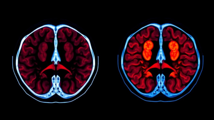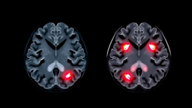
Forensic pathologists have long faced one of the most consequential questions in their discipline: when a person suffers a craniocerebral injury, was the head accelerated into an object, or was it struck and then decelerated? The distinction matters enormously in legal medicine, because it can determine whether a death was an accident, an assault, or something else entirely. Yet traditional biomechanical approaches often falter when injury morphology is atypical, leaving examiners with uncertain conclusions that may be tested in court. A new study published in the International Journal of Legal Medicine by Ya-Wen Liu and colleagues at the Academy of Forensic Science in Shanghai now offers a computational answer, describing a cascaded deep learning system that reads computed tomography images and infers the underlying injury mechanism with remarkable accuracy.
The research team built a two-stage architecture that mirrors how a forensic expert actually works. The first stage performs segmentation: a DeepLabv3+ network, a convolutional model widely used for dense pixel-level prediction, was trained to delineate six distinct injury regions on head CT scans. DeepLabv3+ is particularly suited to this task because of its atrous spatial pyramid pooling module, or ASPP, which applies convolutions at multiple dilation rates in parallel. This allows the network to capture both fine local detail, such as the edges of a small contusion, and broader contextual information, such as the spatial relationship between a hemorrhage and the surrounding skull and brain tissue. In this study, the segmentation network achieved a mean Dice coefficient of 0.87, a standard overlap metric indicating substantial agreement between the model’s injury maps and expert annotations.
The second stage of the cascade handles classification. A ResNet18 backbone, a relatively lightweight residual convolutional network, was combined with the same ASPP module to categorize each case as either an acceleration injury or a deceleration injury. Crucially, the classifier did not operate on raw images alone. Instead, it received segmentation-guided information from the first stage, meaning the features extracted by the segmentation network were fed forward to inform the classification decision. This design choice proved decisive. In an ablation study, in which components of the system are systematically removed to measure their contribution, the macro-F1 score, a balanced measure of precision and recall across classes, improved from 0.71 to 0.82 when segmentation-guided information was included. The cascaded model as a whole attained a mean area under the receiver operating characteristic curve, or AUC, of 0.94 for injury mechanism classification, a figure that places its discriminative power in the range typically associated with expert-level medical imaging systems.
The physics underlying the classification problem explains why it is so difficult. In an acceleration injury, the head is stationary and is set into motion by an impacting force, as when a fist or a blunt object strikes a stationary skull. In a deceleration injury, the head is already moving and is abruptly stopped, as when a falling person’s head strikes the ground or a vehicle occupant’s head hits the windshield. Both scenarios produce contusions, hemorrhages, and fractures, but the distributions of these lesions differ in subtle ways rooted in inertial biomechanics. Coup injuries, arising at the site of impact, and contrecoup injuries, arising on the opposite side of the brain as it rebounds against the inner table of the skull, follow patterns that depend on the direction and magnitude of the forces involved. Distinguishing these patterns reliably has traditionally required expert judgment, and even experts can disagree when the injury presentation is ambiguous.
What makes the new system scientifically interesting is not merely its accuracy but the way its errors and successes align with established biomechanical principles. The authors validated the model externally across frontal, temporal, and occipital regions of the head, and found that its performance patterns were consistent with what biomechanics would predict. This kind of consistency check matters because deep learning models are notorious for exploiting spurious correlations, learning to recognize hospital-specific imaging artifacts or demographic confounds rather than genuine pathology. A model whose regional performance tracks known mechanisms of force transmission is more likely to have learned something real about injury physics, which is essential if the tool is ever to carry weight in a courtroom.
The study also demonstrated that the system could reliably distinguish injured brains from normal images, a prerequisite for any forensic screening application. This binary detection task may seem trivial compared with mechanism classification, but in forensic practice it is far from it. Postmortem changes, varying image quality, and the enormous anatomical variability of human brains all complicate automated analysis. The fact that the cascaded architecture maintained high accuracy on this task while simultaneously performing the harder mechanism classification suggests that the segmentation stage is doing genuine work, localizing pathology in a way that generalizes across cases rather than memorizing individual examples.
The work sits within a rapidly expanding literature on artificial intelligence in forensic imaging. Previous studies have used deep learning to detect skull fractures from curved maximum intensity projections, to identify critical findings in head CT scans, and to classify fractures on the basis of their location and number in order to discriminate between falls and blows. Each of these efforts has attacked a piece of the forensic puzzle. The present study is notable for targeting the mechanism question itself, which is arguably the most legally sensitive piece of all. It also joins a growing family of cascaded and joint segmentation-classification architectures, in which one network’s output refines another’s input, a strategy that has proven effective in medical imaging tasks ranging from cell classification to cancer diagnosis.
The practical implications for forensic practice could be substantial. Determining the manner of injury currently depends on the availability of experienced examiners, and in jurisdictions with heavy caseloads or limited forensic resources, such expertise may be scarce. An automated system that provides an objective, quantifiable assessment of injury mechanism could serve as a second opinion, flagging cases where the imaging evidence contradicts the stated history of events. The authors emphasize that the system offers a route toward objective injury mechanism determination, a phrase that carries particular weight in a field where subjective judgment has historically been the norm. Because all data underlying the findings are freely accessible to other researchers, independent groups can scrutinize and extend the work, which is exactly the kind of transparency forensic science needs.
There are, of course, caveats that temper enthusiasm. The model was trained and validated on data from a single institution, the Academy of Forensic Science in Shanghai, and although external validation across anatomical regions was performed, broader multi-center validation across different scanners, populations, and injury contexts will be needed before deployment. Legal medicine also demands more than a probability score; courts require explainable reasoning, and a 0.94 AUC does not by itself explain why a particular injury pattern indicates deceleration rather than acceleration. Future work will likely need to couple such models with interpretability techniques and with the biomechanical modeling traditions, including finite element analysis, that have long informed the field. The study was approved by the Scientific and Ethical Committee of the Academy of Forensic Science, conducted with informed consent from the families involved, and supported by Chinese national and municipal research programs, reflecting the institutional investment now flowing into computational forensics.
Nevertheless, the study marks a genuine step forward in the marriage of artificial intelligence and legal medicine. By combining a segmentation network that sees where the injury is with a classification network that reasons about how it happened, Liu and colleagues have built a system that approximates, in silicon, the diagnostic chain that forensic experts perform in their heads. The quantitative gains, from a macro-F1 of 0.71 to 0.82 with segmentation guidance, and a mean AUC of 0.94 overall, are not just incremental numbers; they demonstrate that structural information about injury location and extent is the key to unlocking mechanism inference. As such cascaded architectures mature and accumulate validation across centers and populations, the prospect of a forensic imaging tool that can tell investigators not only what happened to a brain, but how it happened, moves from aspiration toward reality, with implications for accident reconstruction, criminal investigation, and the pursuit of justice in cases where the only witness is the injured tissue itself.
Subject of Research: Deep learning classification of acceleration and deceleration craniocerebral injuries for forensic injury mechanism determination
Article Title: Classification of acceleration and deceleration craniocerebral injuries by using cascaded deep learning models
Article References: Liu, Y.-W., Tian, Z.-L., Liu, Y.-Y., Fu, E.-H., Dong, H.-W., Wan, L., Zhang, J.-H., Zou, D.-H., & Liu, N.-G. (2026). Classification of acceleration and deceleration craniocerebral injuries by using cascaded deep learning models. International Journal of Legal Medicine. https://doi.org/10.1007/s00414-026-03971-2
Image Credits: AI Generated
DOI: 10.1007/s00414-026-03971-2
Keywords: craniocerebral injury, forensic medicine, deep learning, DeepLabv3+, ResNet18, image segmentation, computed tomography, injury mechanism, acceleration injury, deceleration injury, biomechanics, legal medicine
Cite Scienmag News
APA MLA Chicago
Cassandra Pierce. (October 3, 2026). Cascaded AI Distinguishes Acceleration From Deceleration Brain Injuries on CT Scans. Scienmag. https://scienmag.com/cascaded-ai-distinguishes-acceleration-from-deceleration-brain-injuries-on-ct-scans/
Cassandra Pierce. “Cascaded AI Distinguishes Acceleration From Deceleration Brain Injuries on CT Scans.” Scienmag, 3 October 2026, https://scienmag.com/cascaded-ai-distinguishes-acceleration-from-deceleration-brain-injuries-on-ct-scans/. Accessed 3 October 2026.
Cassandra Pierce. “Cascaded AI Distinguishes Acceleration From Deceleration Brain Injuries on CT Scans.” Scienmag. October 3, 2026. https://scienmag.com/cascaded-ai-distinguishes-acceleration-from-deceleration-brain-injuries-on-ct-scans/
Copy citation Download RIS
Tags: acceleration injurybiomechanicscomputed tomographycraniocerebral injurydeceleration injurydeep learningDeepLabV3+forensic medicineimage segmentationinjury mechanismlegal medicineResNet18