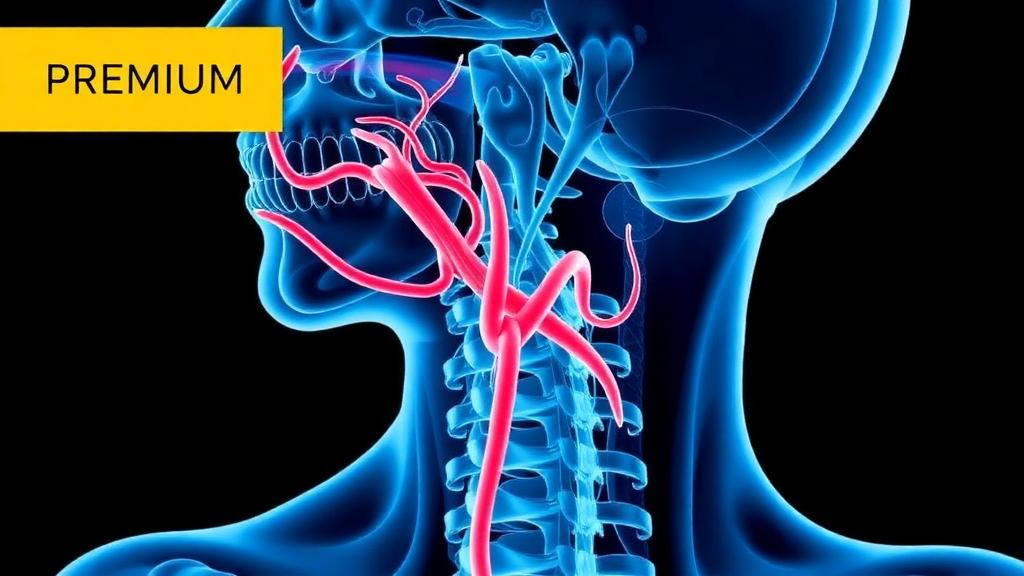
A large cross-sectional study has identified distinct features of carotid artery anatomy that are associated with two different aspects of cardiovascular disease: the presence of arterial narrowing and blood-flow conditions that are potentially favorable to plaque formation. The analysis combined three-dimensional measurements from 346 carotid bifurcations with patient-specific computational fluid dynamics informed by 4D flow MRI for 176 of them. Wider bifurcation and internal carotid artery angles were the strongest geometric markers of stenosis, while the size of the internal carotid bulb and the ratio between vessel areas were more closely associated with low wall shear stress, elevated oscillatory shear and prolonged particle residence in arteries without detectable narrowing. Together, the results suggest that a single anatomical region can carry complementary signals about existing disease and the local flow environment that may precede visible stenosis.
The study was published in the Annals of Biomedical Engineering and analyzed MRI data from 218 people, from whom 240 stenosed and 106 non-stenosed carotid bifurcations were reconstructed. Some participants contributed both sides: among the 128 people with two bifurcations, 47 had different disease status on the left and right. The stenosed group had a mean area-based narrowing of 34.74% with a standard deviation of 16.98 percentage points. Because several patients contributed more than one artery, the researchers also used subject-clustered analyses and cross-validation in which both sides from a person remained in the same data fold. These safeguards did not materially change the main results.
MRI was performed on a 3-tesla clinical scanner using an electrocardiographically triggered, time-resolved three-dimensional phase-contrast sequence. An automated neural-network segmentation system isolated the vessels, achieving a median Dice similarity coefficient of 0.88 on its test set. The processed velocity fields were then used to define inlet and outlet boundary conditions for computational fluid dynamics simulations. These simulations were conducted as quasi-direct numerical simulations with a maximum cell size of 0.07 millimeters, locally refined around stenoses and the carotid bulb, and five near-wall cell layers. Each case covered three cardiac cycles, with the first two discarded so that the reported averages were not dominated by the initial condition. A typical simulation required about 21 hours on 256 processor cores.
To model blood, the investigators used a Newtonian fluid with a viscosity of 0.004 pascal-seconds and a density of 1,060 kilograms per cubic metre. The vessel walls were assumed to be rigid, and the external carotid outlet used a zero-normal-gradient condition, meaning that no specific pressure level or flow rate was imposed there. The internal carotid outlet flow was adjusted to conserve mass while retaining the MRI-derived division of flow between the internal and external carotid arteries. These assumptions are standard simplifications, but they mean that the absolute simulated shear measurements are model-dependent. The authors cited prior estimates indicating that geometric uncertainty can affect carotid wall-shear calculations substantially more than rheology, although rigid and compliant walls can still produce marked differences in peak systolic wall stress.
Among 240 stenosed and 106 non-stenosed bifurcations, several geometric differences were clear. Stenosed arteries had significantly larger bifurcation angles, internal carotid angles, tortuosity and offset flow divider, with adjusted analyses preserving these associations at a p-value below 0.001. The offset flow divider was a new automated descriptor designed to quantify lateral displacement of the actual flow divider from the projected centerline, providing information about apex asymmetry that conventional angles do not directly capture. A four-feature model using bifurcation angle, internal carotid angle, offset flow divider and internal carotid planarity achieved a cross-validated area under the receiver operating characteristic curve, or AUROC, of 0.767, with a 95% confidence interval of 0.716 to 0.818. Its recall was 0.90 for stenosed cases but only 0.39 for non-stenosed cases, confirming that the model is not a ready-made diagnostic test.
The stenosis label itself was assigned from morphometric criteria applied to the same segmented three-dimensional surfaces used for the geometric measurements. One mechanism identified reduced cross-sectional roundness, with a threshold of 0.95, while another detected a relative change of at least 3% per millimetre along a branch. This shared imaging foundation creates an important limitation: the classification and anatomical features are not fully independent, and the study demonstrates association with prevalent stenosis rather than the ability to forecast future narrowing. The modest discrimination also reflects an imbalance in the cohort, since 69.4% of the bifurcations were stenosed. In a balanced version of the model, sensitivity was 0.76 and specificity 0.67, with no improvement in overall discrimination.
Patient-specific simulations in 101 stenosed and 75 non-stenosed bifurcations revealed a more detailed relationship between anatomy and flow. The degree of narrowing itself correlated only weakly with the post-stenotic hemodynamic indices: the correlations were 0.09 for time-averaged wall shear stress, 0.28 for the oscillatory shear index and -0.13 for relative residence time. By contrast, the ratio describing expansion immediately beyond the stenosis was associated with stronger disturbance. In moderate stenoses, expansion correlated with the oscillatory shear index at r = 0.80 and with the backflow index at r = 0.77. The association weakened among severe cases, although the researchers did not formally test whether correlations differed between severity groups and noted that estimates in the smallest subgroup were sensitive to one influential case and to acquisition differences.
The clearest result emerged in the 75 non-stenosed bifurcations. After the measured outcomes were normalized by each participant’s mean common carotid flow rate, models based on seven geometric features explained 57.5% of the variation in minimum time-averaged wall shear stress, 60.3% in maximum relative residence time and 49.0% in maximum oscillatory shear. Bulb area was the largest independent correlate of low wall shear and long residence, uniquely accounting for 22.6% and 21.2% of their variance, respectively. Area ratio was the strongest independent correlate of oscillatory shear, with a unique contribution of 18.2%. A composite ranking of internal carotid angle, bulb area and area ratio correlated with the combined low-shear, high-oscillation and long-residence profile at Spearman rho = 0.750, although this was an exploratory within-sample result rather than independent validation.
Representative cases illustrated the contrast: four non-stenosed bifurcations with large bulbs had relative residence times of roughly 7 to 17, compared with about 0.7 to 1.1 in four cases with small bulbs. Their wall maps showed the largest low-shear and high-oscillation regions along the outer wall of the internal carotid artery, a location commonly associated with plaque initiation. The findings are not evidence that a particular individual will develop plaque, nor do they establish that the measured flow patterns cause atherosclerosis. The cross-sectional design cannot determine whether abnormal geometry created the flow disturbance, plaque remodeling produced both altered angles and stenosis, or both processes contributed. The cohort was older, lacked external validation, did not model plaque composition, and drew from two MRI acquisition protocols with different velocity-encoding settings. Despite these constraints, the analysis identifies measurable anatomical variables for longitudinal testing: the structure of the carotid bulb may reveal a disturbed-flow environment even before narrowing becomes apparent, while post-stenotic expansion may describe the local flow burden more fully than narrowing percentage alone.
Subject of Research: Associations between carotid bifurcation geometry, stenosis, and simulated local hemodynamics
Article Title: Hemodynamic and Geometrical Parameters Associated with Carotid Artery Stenosis: A Cross-Sectional Study Using 4D Flow MRI and CFD
Article References: Mokhtari, A., Bergmann, A., Strecker, C., Andrae, J., Harloff, A., & Obrist, D. (2026). Hemodynamic and Geometrical Parameters Associated with Carotid Artery Stenosis: A Cross-Sectional Study Using 4D Flow MRI and CFD. Annals of Biomedical Engineering. https://doi.org/10.1007/s10439-026-04417-x
Image Credits: AI Generated
DOI: 10.1007/s10439-026-04417-x
Keywords: carotid stenosis, 4D flow MRI, computational fluid dynamics, arterial geometry, wall shear stress, atherosclerosis, blood flow, carotid bifurcation, medical imaging, Hemodynamic, Geometrical, Parameters
News Source: Ophelia Keating. (October 19, 2026). Carotid Geometry Links to Stenosis and Disturbed Blood Flow. Scienmag.