A rare presentation of life-threatening cardiac tamponade resulting from effusive-constrictive pericarditis developed in a seventeen-year-old male living with common variable immunodeficiency, according to a detailed clinical case report published in the International Journal of Emergency Medicine. The young patient, who had been diagnosed with the underlying primary immunodeficiency disorder in 2022, arrived at an emergency department suffering from severe hypoxemia, generalized weakness, dry cough, and marked dyspnea that had progressed over several days prior to admission. While cardiovascular manifestations remain exceedingly uncommon among individuals diagnosed with primary antibody deficiencies, this clinical presentation highlights an intricate pathophysiological scenario wherein fluid accumulation in the pericardial space acutely compressed the myocardium, generating hemodynamic compromise that subsequently unmasked an underlying constrictive physiology once the fluid was drained. Emergency clinicians and cardiologists were confronted with an acute diagnostic and therapeutic challenge requiring immediate invasive decompression alongside an extensive multidisciplinary diagnostic workup to exclude infectious, tuberculous, and malignant etiologies in an immunocompromised host.
Common variable immunodeficiency represents one of the most frequently identified symptomatic primary immunodeficiencies globally, carrying an estimated general prevalence ranging from approximately one in 25,000 to one in 50,000 individuals. Pathophysiologically, the disorder is characterized by substantially decreased circulating immunoglobulin levels and impaired specific antibody production, which predisposes affected individuals to recurrent bacterial and opportunistic infections predominantly localized within the respiratory and gastrointestinal tracts. Beyond infectious susceptibility, modern immunological literature increasingly emphasizes that widespread immune dysregulation forms a substantial pathophysiological axis of the disease spectrum. In large prospective and retrospective cohort investigations encompassing 473 patients, non-infectious inflammatory complications occurred in approximately two-thirds of all cases and were strongly linked to elevated morbidity and mortality. These extra-infectious manifestations include autoimmune cytopenias, organ-specific autoimmune diseases, enteropathies, chronic pulmonary remodeling, non-caseating granulomatous inflammation, lymphoid hyperplasia, and an elevated baseline susceptibility to lymphoproliferative malignancies.
The medical history of the seventeen-year-old patient reflected a lifelong burden of systemic illness directly attributable to severe antibody deficiency and disordered immune homeostasis. Baseline immunological quantification confirmed profound hypogammaglobulinemia, with serum immunoglobulin G measured at 180 milligrams per deciliter, serum immunoglobulin A falling below 10 milligrams per deciliter, and serum immunoglobulin M measured at 15 milligrams per deciliter. To manage his immune deficiency, the patient received regular maintenance therapy comprising monthly infusions of intravenous immunoglobulin at a dose of five grams, with his last administration occurring eighteen days prior to the acute emergency presentation. His complex past history included recurrent lower respiratory tract infections, diagnosed inflammatory bowel disease, biopsy-confirmed celiac disease, a documented episode of malaria, a prior right-sided psoas abscess, an internal posterior anal fissure, and first-degree internal hemorrhoids. Furthermore, chronic malabsorption and recurring systemic illness caused severe nutritional wasting, leaving the adolescent with a body weight of merely 25 kilograms and a severely diminished body mass index of 15 kilograms per square meter.
Upon initial physical examination in the emergency ward, the patient displayed unmistakable evidence of critical cardiopulmonary distress and profound hemodynamic compromise. Ambient room air pulse oximetry registered a critically low peripheral oxygen saturation of 75 percent, which improved to 100 percent only after the immediate administration of supplemental oxygen delivered via nasal cannula at three liters per minute. Hemodynamic parameters revealed resting sinus tachycardia at 88 beats per minute, tachypnea at 27 breaths per minute, a normothermic core body temperature of 37 degrees Celsius, and systemic arterial hypotension registering 90/60 millimeters of mercury. Classical physical findings associated with Beck’s diagnostic triad were evident, including marked engorgement and visible distension of the neck veins reflecting elevated jugular venous pressure, alongside muffled, distant heart sounds on precordial auscultation. A pronounced pulsus paradoxus, quantified as an inspiratory reduction in systolic blood pressure exceeding ten millimeters of mercury, was readily detected, raising immediate clinical suspicion for cardiac tamponade, while chest auscultation demonstrated bilateral basal and mid-zone inspiratory crackles accompanied by bilateral dependent pitting pedal edema.
Initial laboratory evaluations demonstrated marked systemic inflammation alongside severe hypoalbuminemia indicative of protein malnutrition and chronic catabolism. Peripheral blood tests identified an elevated serum C-reactive protein concentration of 23 milligrams per liter and a total white blood cell count of 12,400 cells per microliter with absolute neutrophilia, alongside a hemoglobin concentration of 9.8 grams per deciliter and a platelet count of 310,000 per microliter. Biochemical markers of hepatic and renal function remained preserved, demonstrating blood urea nitrogen of 24 milligrams per deciliter, serum creatinine of 0.7 milligrams per deciliter, total serum bilirubin of 0.8 milligrams per deciliter, alanine transaminase of 28 units per liter, and aspartate transaminase of 34 units per liter. However, total serum protein was notably depressed at 4.05 grams per deciliter, and serum albumin measured only 1.8 grams per deciliter. Comprehensive contrast-enhanced computed tomography of the neck, chest, abdomen, and pelvis confirmed circumferential pericardial effusion and active pericarditis, accompanied by bilateral ground-glass pulmonary opacities, mild ascites, hepatosplenomegaly, and diffuse mesenteric and hilar lymphadenopathy without evidence of necrotic mediastinal nodes or pulmonary cavitary lesions.
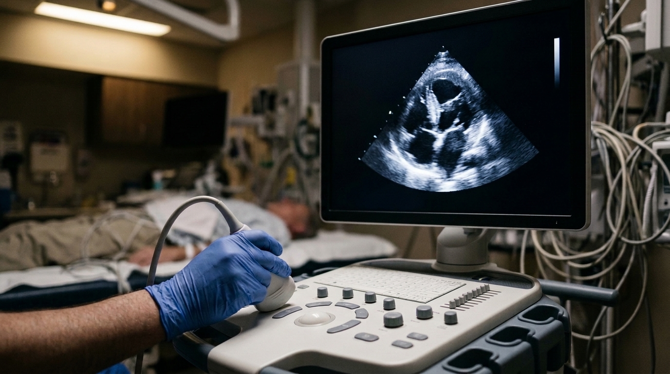
Transthoracic echocardiography performed at the bedside proved vital in characterizing the pericardial space and documenting severe abnormal ventricular interaction. Imaging demonstrated a moderate circumferential pericardial effusion measuring twelve to fifteen millimeters in anterior-posterior diameter, containing a prominent, dense, highly mobile echogenic mass highly characteristic of an organized intrapericardial fibrin clot. While overt diastolic collapse of the thin-walled right ventricle was not observed, late diastolic inward motion of the right atrial free wall was clearly identified.
Given the combination of worsening hypotension, elevated venous pressures, pulsus paradoxus, and echocardiographic signs of physiological compromise, the medical team executed urgent diagnostic and therapeutic pericardiocentesis. A pigtail catheter inserted under echocardiographic guidance remained in place across a 48-hour drainage period, successfully evacuating a cumulative volume of 320 milliliters of turbid, light yellow-to-brown serosanguineous fluid containing macroscopic fibrinous clots. Quantitative fluid analysis revealed a nucleated cell count of 16,000 cells per cubic millimeter with 85 percent mature lymphocytes and 15 percent polymorphonuclear leukocytes, alongside a red blood cell count of 6,400 cells per cubic millimeter. Biochemical assessment established a pericardial fluid protein level of 1.36 grams per deciliter against a concurrent serum protein level of 4.05 grams per deciliter, yielding a ratio of 0.34, whereas pericardial fluid lactate dehydrogenase was profoundly elevated at 10,348 units per liter against a serum level of 502 units per liter, generating an LDH ratio of 20.61 that firmly satisfied Light’s criteria for an inflammatory exudate.
Exhaustive pathological and microbiological evaluations were immediately conducted to differentiate between infectious, tuberculous, autoimmune, and neoplastic triggers, particularly because the patient resided in a geographic region where Mycobacterium tuberculosis remains endemic. Cytological smears and cell-block preparations demonstrated dense lymphocytic inflammatory aggregates mixed with organized fibrin networks but confirmed a total absence of malignant or atypical cells, confirming that the mobile intrapericardial mass was an organized inflammatory clot rather than an intracardiac tumor. Direct acid-fast bacilli staining using the Ziehl-Neelsen technique, real-time polymerase chain reaction via the Xpert MTB/RIF assay, and prospective mycobacterial cultures of both pericardial aspirate and sputum samples were uniformly negative, with fluid adenosine deaminase testing showing non-elevated concentrations. Furthermore, Gram stains and bacterial fluid cultures remained sterile, three independent sets of peripheral blood cultures yielded no growth, and targeted molecular viral assays excluding active infection from Coxsackievirus B, echovirus, cytomegalovirus, Epstein-Barr virus, and SARS-CoV-2 returned negative results, leaving an immune-mediated inflammatory mechanism as the most plausible primary driver.
The patient was stabilized through a structured medical management strategy incorporating broad-spectrum antimicrobial coverage, hemodynamic support, targeted nutritional rehabilitation, and serial physiological monitoring. Empirical intravenous therapy consisting of meropenem at one gram every eight hours and vancomycin at 500 milligrams every twelve hours was administered for ten days, complemented by inhaled bronchodilators, inhaled corticosteroids, and intravenous tranexamic acid at 500 milligrams twice daily during initial drainage to mitigate potential hemorrhagic progression within the fibrin-rich space. To treat severe hypoalbuminemia, salt-free twenty percent human albumin was infused at 100 milliliters daily for three days alongside therapeutic probiotics and targeted enteral nutrition. Although drainage of the fluid rapidly alleviated overt clinical tamponade and normalized peripheral oxygenation, central venous pressure unexpectedly remained elevated between 14 and 16 millimeters of mercury. Repeat transthoracic echocardiography on the fourth post-procedure day confirmed complete fluid clearance and an improved ejection fraction of 52 percent, yet demonstrated persistent Doppler signs of constrictive physiology, including greater than 25 percent respiratory variation in mitral inflow velocity, prominent hepatic vein diastolic flow reversal during expiration, a plethoric inferior vena cava, and marked septal shudder, conclusively confirming effusive-constrictive pericarditis.
Remarkably, sustained conservative management facilitated the gradual and complete resolution of this constrictive hemodynamic state without requiring surgical intervention or formal subtotal pericardiectomy. Continued nutritional support and regular replenishment of intravenous immunoglobulin contributed to clinical recovery, with systemic inflammatory markers normalizing as serum C-reactive protein fell dramatically from 23 milligrams per liter to four milligrams per liter by discharge on hospital day twelve. At a scheduled three-month outpatient follow-up evaluation, the adolescent remained entirely asymptomatic, reporting no recurrence of dyspnea, fatigue, or chest discomfort, while nutritional rehabilitation increased his serum albumin concentration to 3.2 grams per deciliter. Follow-up transthoracic echocardiography revealed preserved biventricular systolic performance, physiological Doppler filling dynamics without residual constrictive patterns, and complete resolution of the previously visualized mobile intrapericardial fibrin mass. The authors conclude that cardiopulmonary deterioration in immunocompromised patients must prompt consideration of primary cardiac etiologies alongside routine pulmonary infections, underscoring the necessity of a two-step diagnostic paradigm that first relieves mechanical tamponade and subsequently reassesses intracardiac filling dynamics to identify transient effusive-constrictive pathology.
Subject of Research: Clinical presentation and non-surgical resolution of effusive-constrictive pericarditis causing cardiac tamponade in common variable immunodeficiency
Article Title: Cardiac tamponade caused by effusive-constrictive pericarditis in a patient with common variable immunodeficiency: a case report
Article References: Khan, A., Tahir, O., Batool, A., Farman, J., Javaid, M. H., & Farhaan, A. R. (2026). Cardiac tamponade caused by effusive-constrictive pericarditis in a patient with common variable immunodeficiency: a case report. International Journal of Emergency Medicine, 19(1), Article 243. https://doi.org/10.1186/s12245-026-01381-0
Image Credits: AI Generated
DOI: 10.1186/s12245-026-01381-0
Keywords: Common Variable Immunodeficiency, Effusive-Constrictive Pericarditis, Cardiac Tamponade, Pericardiocentesis, Hypogammaglobulinemia, Echocardiography, Pulsus Paradoxus, Intravenous Immunoglobulin, Pericardial Effusion, Central Venous Pressure, Septal Bounce, Mitral Inflow Variation, Exudative Effusion, Light's Criteria, Immune Dysregulation, Plethoric Inferior Vena Cava, Ventricular Interdependence
News Source: Ophelia Keating. (November 19, 2026). Cardiac Tamponade from Effusive Constrictive Pericarditis Arises in Common Variable Immunodeficiency. Scienmag.