


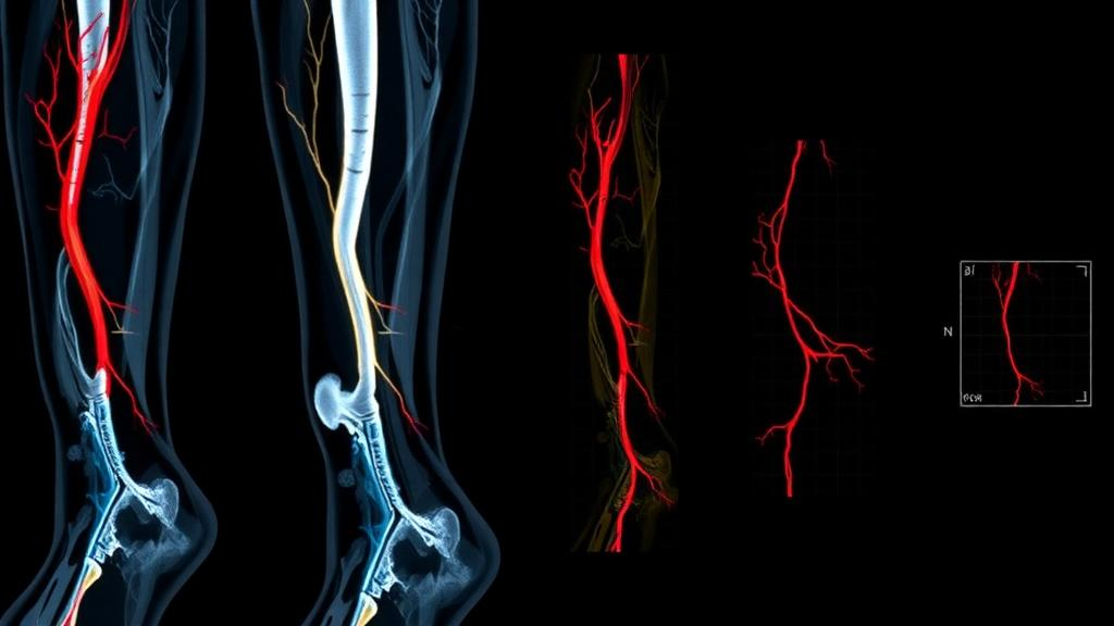
Researchers have found that the texture of calf muscles, as seen on computed tomography angiography (CTA), is associated with the severity of lower-limb arterial disease in patients with peripheral arterial disease. The retrospective study, published in BMC Medical Imaging, suggests that a purely image-based measurement of muscle appearance could complement existing assessments of how badly arteries in the leg are affected.
The study asked a different question: whether directional texture features extracted from the posterior calf muscles on CTA images carry information about that severity, without requiring new measurements of the vessels themselves.
The research team analysed 30 patients with peripheral arterial disease, covering 60 lower limbs. From dynamic CTA scans, they selected six consecutive axial slices centred on the maximum posterior calf muscle cross-sectional area, taken at both the 15-second and 30-second phases of the scan. The gastrocnemius and soleus muscles were segmented, and 70 grey-level co-occurrence matrix texture features were extracted for each phase, capturing directional patterns in how pixel intensities are arranged within the muscle tissue.
To avoid overfitting, the researchers built phase-specific L1-penalized logistic regression models, using patient-grouped cross-validation and the one-standard-error rule for feature selection. The final 1-SE LASSO models retained 10 features at 15 seconds and 9 features at 30 seconds.
Performance was assessed with internal validation using 500 patient-cluster bootstrap resamples, with the entire model-development procedure repeated within each resample. After bootstrap correction for optimism, the area under the curve was 0.769 for the 15-second model and 0.695 for the 30-second model. The 15-second model showed numerically higher discrimination, although the authors note that no formal comparison between the two phases was performed.
The authors conclude that CTA-based directional texture features of the posterior calf muscles were associated with runoff score-defined lower-limb arterial disease severity, and that these findings support further evaluation of directional calf-muscle texture analysis in larger independent cohorts. As a retrospective study of 30 patients with internal validation only, the results are preliminary and require external confirmation before any clinical use.
Subject of Research: CTA-based directional texture analysis of calf muscles in peripheral arterial disease severity assessment
Article Title: CTA-based directional texture analysis of calf muscle in peripheral arterial disease: association with runoff score–defined lower-limb arterial disease severity
Article References: Wei, X., Lin, Z., Zhang, D., & Zhou, X. (2026). CTA-based directional texture analysis of calf muscle in peripheral arterial disease: association with runoff score–defined lower-limb arterial disease severity. BMC Medical Imaging. https://doi.org/10.1186/s12880-026-03016-5
Image Credits: AI Generated
DOI: 10.1186/s12880-026-03016-5
Keywords: peripheral arterial disease, CTA, texture analysis, calf muscle, runoff score, radiomics, LASSO, logistic regression, bootstrap validation, BMC Medical Imaging, CTA-based, directional
News Source: Ophelia Keating. (October 29, 2026). Calf muscle texture patterns on CT scans track artery disease severity. Scienmag.



