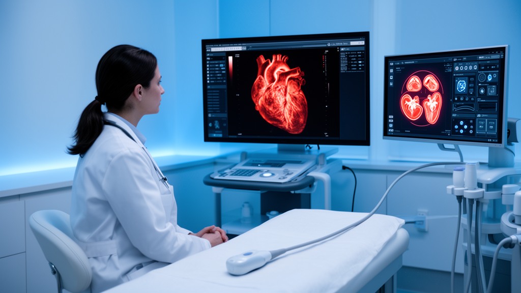
Artificial intelligence is poised to reshape nearly every stage of the echocardiogram, from booking the appointment to interpreting the final images, but the British Society of Echocardiography warns that clinical adoption remains limited and uneven despite an exponential expansion of research in the field. In a new position statement published in Echo Research & Practice, the society offers a structured, consensus-driven evaluation of the opportunities, limitations and requirements for integrating AI into echocardiography services safely, equitably and effectively. The document was developed by a multidisciplinary writing group of cardiologists, cardiac physiologists and clinical scientists, clinical academics, AI researchers, and patient and public representatives, with consensus reached through iterative discussion, structured review rounds and final approval by the BSE Trustees. Although centred on the United Kingdom, the society notes that many of its principles may be relevant to other healthcare systems with similar models of echocardiography delivery.
The statement is deliberately not a traditional narrative review. Instead, it articulates the BSE’s position across the entire echocardiography workflow, informed by a systematic review of the literature registered on PROSPERO and by input from patients and the public. To convey how mature each AI application actually is, the writing group adopted the technology readiness level scale, a nine-level framework widely used to estimate the developmental stage of new technologies. The scale allows a clear distinction between early-stage models that exist only in the laboratory and those that have gained regulatory approval, funding approval and deployment in real-world healthcare settings. The authors used this framework to separate AI tools that are ready for clinical deployment from those that still require further evidence, and they stress that because the field is evolving so rapidly, the consensus will require regular review and, where needed, an updated position statement will follow.
At the front end of the pathway, the society sees realistic promise in referral, triage and scheduling. Deep learning models have been shown to support accurate and efficient triage of appropriate referrals, and large language models have the potential to support referrals and triage by integrating patient information, clinical presentation and societal triage guidelines, potentially enabling prioritisation of patients based on clinical need. AI may also predict peak attendance times and identify demographics of patients less likely to attend at certain times, allowing optimal scheduling and service efficiency. However, the statement notes there is currently a lack of AI models focusing on scheduling within echocardiography, placing such tools at the lowest readiness levels. The patient interaction that echocardiographers provide before, during and after the scan, including checking the appropriateness of referrals and explaining findings, is unlikely to be replaced by AI.
Image acquisition is one area where AI has already reached clinical maturity. Echocardiography is a specialised modality requiring highly trained echocardiographers, and acquiring a comprehensive, standardised imaging dataset within a limited time frame can be particularly challenging for inexperienced operators, especially in emergency or community settings where access to expertise is limited. AI models have been shown to improve the acquisition of correct and diagnostic images, which may support the training of novice echocardiographers and aid earlier diagnosis and timely treatment where experienced staff are scarce. This technology is currently available across a range of echocardiographic vendors and sits at the top of the readiness scale. By contrast, AI models designed to improve image quality itself, such as deep learning approaches that identify and remove noise and artefacts, and work on fusing multiple 3D images with breath holds so the myocardium is displayed in the same position across acquisitions, remain within the research and development phase.
Quantitative analysis is perhaps the most mature application of all. Echocardiography is operator-dependent, and despite well-established guidelines, intra- and inter-operator variability remains a significant challenge affecting the reliability and reproducibility of measurements. The statement highlights left ventricular ejection fraction as a prime example, with previous studies demonstrating inter-observer variability of up to plus or minus 14 per cent; for patients receiving cardiotoxic chemotherapy, such variability may result in misjudgement of ejection fraction and potentially unnecessary treatment delays. AI models are increasingly demonstrating the ability to standardise measurements and reduce variability, particularly for ejection fraction, left ventricular global longitudinal strain, valvular heart disease assessment and right ventricular chamber quantification. Automated measurements of cardiac structure and function are now commonplace and available on most up-to-date echocardiography machines, while the majority of other quantification models remain in the research or development phase, with few having gained regulatory approval.
Beyond measurement, AI is showing promise in detecting disease states that may be beyond human perception by analysing features from images, tabular data, or both in combination. The statement lists promising applications including detection of coronary artery disease on stress echocardiography, identification of cardiac amyloidosis, characterisation of intracardiac masses, and determining the aetiology of left ventricular hypertrophy, where AI models have been able to accurately and reliably distinguish between athlete’s heart, hypertrophic cardiomyopathy and hypertensive heart disease. AI has also shown utility in assessing valvular heart disease, including aortic stenosis and mitral, tricuspid and aortic regurgitation. Several models have accumulated sufficient evidence to gain regulatory approval, while others remain in earlier stages of development. In risk stratification, AI models may identify previously unknown echocardiographic markers or imaging phenotypes; in aortic stenosis, a model combining echocardiography, cardiac magnetic resonance and computed tomography data with blood test results was able to identify subgroups of patients at higher risk of poor clinical outcomes, potentially improving the timing of valve interventions. These risk models, however, remain in the research and development phase.
The society is notably cautious about productivity claims. Studies have demonstrated that AI assistance can reduce echocardiography time, fuelling expectations of higher patient throughput amid rising demand and persistent workforce constraints, and AI-enabled reductions in scanning time may lessen the ergonomic burden on echocardiographers by shortening prolonged static postures that contribute to work-related musculoskeletal disorders. Yet the benefits are not universal: in cases with fair or poor image quality, AI tools require more corrections and longer measurement times compared with high-quality studies. There remains limited real-world evidence from NHS settings to determine whether AI can meaningfully increase patient activity, particularly since many centres already have access to built-in automation within existing platforms and continue to operate with 45 to 60 minute appointment timeslots. Claims of productivity gains must be interpreted cautiously until robust, context-specific data is available, the statement concludes, and any efficiencies achieved must not come at the cost of patient interaction, clinical interpretation or quality assurance.
The statement also confronts workforce and governance risks head-on. Emerging evidence suggests that exposure to AI models can negatively influence clinician behaviour, potentially contributing to a decline in care standards, and when AI outputs are perceived as authoritative, automation bias may erode critical thinking, leading professionals to overlook errors. Overreliance can develop if operators are not required to remain actively engaged in supervising algorithms, a phenomenon described as operator’s dropout, while the accessibility of AI tools may foster unwarranted confidence among professionals with limited subject-matter expertise. AI-guided acquisition could enable individuals with limited training to obtain diagnostic images, expanding access in underserved settings but risking examinations performed by operators unable to recognise suboptimal windows, artefacts or clinically significant abnormalities. The society’s proposed solutions centre on human-AI collaboration, quality assurance, retention of healthcare professional oversight, and ongoing professional development, with AI treated as an augmentation tool whose final interpretation and decision-making remain with clinicians, supported by new competencies in data literacy, AI model interpretation and quality assurance.
On transparency and ethics, the authors argue that accurate, reproducible and clinically useful models depend fundamentally on the quality, completeness and diversity of training datasets. Certain patient groups, including women, ethnic minorities, and individuals with congenital heart disease or valve repairs and replacements, are often underrepresented, and models are typically trained, tested and validated on non-UK populations, which can exacerbate health inequalities and cause underperformance for underserved groups. The statement calls for training and validation datasets that reflect the diversity of real-world patients, particularly within UK populations and NHS settings, and notes that performance metrics can be misleading when disease prevalence is low, with area under the precision-recall curve offering a more informative assessment in imbalanced datasets. Regulatory pathways also differ: in the United States, most AI-enabled models are cleared through the FDA 510(k) pathway, which does not require prospective clinical evidence of safety, effectiveness or generalisability, and there is currently no clear consensus on how liability should be assigned when AI contributes to a diagnosis or treatment resulting in patient harm. The society also flags infrastructure as a bottleneck, noting that many AI methods require images to be uploaded to off-site cloud environments, introducing dependencies on secure, high-bandwidth connectivity and clear data-transfer agreements.
Patients and the public were directly involved in shaping the statement through group discussions and one-to-one interviews with members of the Stafford and Stoke-on-Trent Heart Support Group and the Leeds Cardiovascular PPIE group. Participants expressed strong support for AI where it could assist healthcare professionals and improve care, but insisted the technology should enhance rather than replace staff. They raised concerns about whether all staff would have the skills to interpret and act on AI-generated results, about equity of access across the NHS, about the reliable transfer of AI-generated data into electronic health records, and about the security of patient data, with several participants also expressing concern about the environmental impact of increased energy usage. Looking forward, the BSE identifies future opportunities including automated image stacking, standardised comparison of serial studies, integrated audit and quality-improvement tools, and real-time flagging of measurement or reporting inconsistencies before report sign-off. The society commits to webinars, e-learning modules and practical workshops for its members, and to considering the incorporation of AI models into future BSE guidance where supported by robust evidence, so that AI is adopted safely, transparently and in a way that improves patient care across all echocardiography services.
Subject of Research: A consensus position statement on the opportunities, limitations and requirements for integrating artificial intelligence into echocardiography services.
Article Title: Artificial intelligence in echocardiography: a position statement from the British Society of Echocardiography
Article References: Bennett, S., Wild, C., Paton, M. F., Victor, K., Khan, S., Kirkham, J., Gaye, G., Peacock, C., Robinson, S., Manisty, C., Muraru, D., Leeson, P., Sengupta, P. P., Augustine, D. X., & On behalf of Stafford and Stoke on Trent Heart Support Group (2026). Artificial intelligence in echocardiography: a position statement from the British Society of Echocardiography. Echo Research & Practice, 13(1), Article 26. https://doi.org/10.1186/s44156-026-00124-4
Image Credits: AI Generated
DOI: 10.1186/s44156-026-00124-4
Keywords: artificial intelligence, echocardiography, British Society of Echocardiography, position statement, cardiac imaging, machine learning, clinical governance, health equity, workforce, regulatory approval, patient and public involvement, NHS
News Source: Blake Davidson. (October 29, 2026). British Society of Echocardiography Sets Out How AI Should Enter the Heart Scan Workflow. Scienmag.