A brief, easily missed seizure type in which a child’s arm suddenly drops or a held object slips from the hand has been mapped in detail by a team studying 17 children treated at a single Chinese hospital. The condition, epileptic negative myoclonus, is caused not by a muscle jerk but by the opposite: a split-second shutdown of muscle activity driven by abnormal electrical discharges in the brain’s sensorimotor cortex. Because the episodes last only a few hundred milliseconds and often leave no obvious trace to an observer, they are frequently overlooked, delaying diagnosis in children who may already be receiving treatment for other forms of epilepsy.
The new report, published in Acta Epileptologica, is a retrospective descriptive analysis of all consecutive patients diagnosed with epileptic negative myoclonus at Wuxi Children’s Hospital between October 2017 and June 2024. The 17 children accounted for 1.28 percent of the 1,323 epilepsy patients seen at the department during that window. There were five boys and twelve girls, a marked female predominance that the authors flag as a potentially important clue about the condition’s underlying biology. The average age at which epilepsy first appeared was 5.71 years, with a range from 3.7 to 9.0 years, and the median interval between the first epilepsy symptom and the emergence of negative myoclonus was eight months, although in some children the two coincided and in others the gap stretched to 33 months.
Epileptic negative myoclonus has a relatively short history as a formally recognized entity. It was first described in 1993, when researchers used simultaneous electroencephalogram and electromyogram recordings to show that transient loss of postural tone was time-locked to epileptiform discharges arising from the contralateral sensorimotor cortex. The International League Against Epilepsy classified it as an independent seizure type in 2001, and in its 2025 classification update the organization explicitly placed it within the framework of motor seizures. Clinically, the episodes can appear as head nodding, hand tremor, intermittent upper limb weakness, unstable grasping, or limb drooping; when the lower limbs are involved, children may become unsteady while standing or fall.
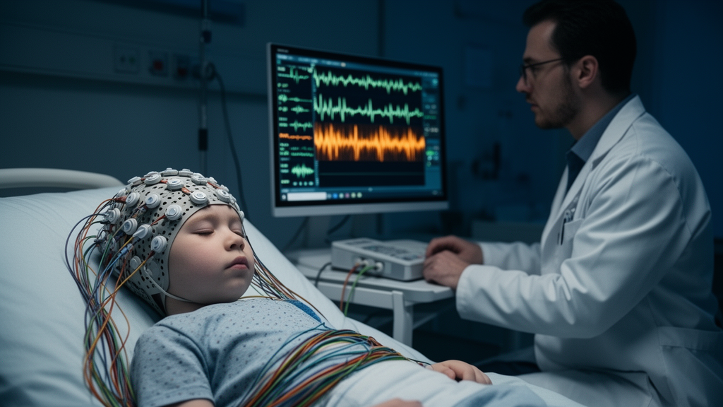
To capture such fleeting events, the investigators relied on a standardized provocation procedure. During long-term video-electroencephalographic monitoring, every child underwent an orthostatic arm extension test: standing awake with both arms held horizontally, the child was observed for transient drooping, nodding, or unstable stance, while synchronized electromyography recorded activity from the deltoid and quadriceps muscles. The test successfully elicited typical events in all 17 children. The recorded episodes consisted of brief drooping of one or both upper limbs, synchronized with slow spikes over the contralateral or bilateral centro-parietal regions. Electromyography showed a silent period in the deltoid muscle lasting 50 to 200 milliseconds, with a mean of 100.59 milliseconds, and no preceding muscle burst, confirming the time-locked relationship between the electrical discharge and the muscle silence. Two board-certified EEG neurologists independently reviewed the recordings, reaching a Cohen’s kappa of 0.92, which the authors describe as excellent agreement.
A striking topographic pattern emerged from the recordings. Upper limb episodes were consistently associated with discharges over the contralateral central and parietal regions, while the two children whose lower limbs were affected showed ictal slow spikes in midline regions, at electrode positions Cz and Pz. This somatotopic correspondence, in which the location of the electrical discharge predicts which limb loses tone, matches the organization of the sensorimotor cortex and is consistent with earlier larger case series. The researchers also found that the spike-and-wave complexes occurring during the episodes had higher frequency and amplitude than interictal discharges, a difference that reached statistical significance, and they suggest this may reflect an irritative phenomenon within the epileptic network.
The study also documented a close relationship with a sleep-related electrographic pattern. Thirteen of the 17 children, or 76 percent, showed a spike-wave index exceeding 60 percent in the rolandic region during non-REM sleep, the operational threshold the team used to identify electrical status epilepticus during sleep. Nine of these children were diagnosed with epileptic encephalopathy with spike-and-wave activation in sleep, a condition defined by cognitive or behavioral regression accompanying that electrographic pattern; all nine presented with clear evidence of neurocognitive or functional deterioration. Among the 12 children in the cohort who had self-limited epilepsy with centrotemporal spikes, negative myoclonus emerged during antiseizure medication therapy after an initial presentation of focal seizures alone, and ten of those 12 developed the sleep-related pattern.
Brain imaging was normal in 16 of the 17 children, but one case proved pivotal. That child presented with negative myoclonus as the initial symptom, with right upper limb drooping during the arm extension test, left central-parietal spike-and-wave complexes, and the sleep-related electrical pattern. Magnetic resonance imaging and PET/CT both suggested focal cortical dysplasia, and after surgical resection of the epileptogenic lesion at Peking University First Hospital in May 2023, histopathology confirmed focal cortical dysplasia type IIa. During nearly two years of follow-up the child remained seizure-free and discontinued all antiseizure medications, with no cognitive or language deficits, although a mild residual motor deficit persisted, noticeable mainly in fine motor skills during high-intensity activities.
Treatment outcomes across the cohort varied considerably. All 17 children initially received antiseizure medication monotherapy, with levetiracetam chosen for eight, valproate for seven, oxcarbazepine for one, and lacosamide for one. One child who started on oxcarbazepine developed negative myoclonus and dysarthria, and switching to valproate produced good control, reinforcing the authors’ caution that oxcarbazepine may induce or aggravate this seizure type. Two children achieved lasting seizure freedom on monotherapy, and four more became seizure-free on dual therapy, three with added clonazepam and one with a levetiracetam-valproate combination. Nine children, however, failed two or more medications and developed the sleep-related electrical pattern with cognitive decline; they received a six-month course of monthly methylprednisolone pulse therapy. Four of the nine responded well, becoming seizure-free with spike-wave index reductions of 30 to 55 percent and cognitive gains of 12 to 23 points. Four others improved initially but relapsed roughly six months after steroid withdrawal, prompting a second course in two of them, both of whom then achieved sustained control. The remaining child responded poorly to steroids but improved markedly after starting a ketogenic diet, remaining seizure-free for 1 year 4 months with an 85 percent reduction in the spike-wave index and a 25-point cognitive gain.
The median follow-up for the whole cohort was 40 months, ranging from 21 to 79 months, and every child was followed for more than a year. The authors emphasize that their findings are hypothesis-generating rather than definitive. The retrospective design, the requirement for available brain MRI data, the modest sample size, the lack of genetic testing in most patients, and the absence of systematic long-term neuropsychological follow-up all limit the strength of the conclusions, and the durability of the cognitive improvements observed after steroid therapy beyond six months remains uncertain. The team also notes that ethosuximide, a drug sometimes considered for this condition, was not used because it was not available locally.
Despite these constraints, the report offers practical guidance for clinicians encountering children with unexplained brief lapses in limb control. The authors recommend early EEG and neuroimaging to improve diagnostic accuracy, suggest that the orthostatic arm extension test be considered when sleep-related electrical activity is present, and advise that children with negative myoclonus and the sleep-related pattern who respond poorly to medication undergo comprehensive evaluation for structural causes such as cortical dysplasia. They also note that isolated negative myoclonus, seen as the sole seizure type in two children in this cohort, may be an early indicator of subsequent development of the sleep-related encephalopathy, arguing that recognition of this pattern should prompt timely sleep EEG monitoring and intervention. Larger, prospective, multicenter studies with standardized cognitive assessment will be needed to confirm these observations in more diverse populations.
Subject of Research: Clinical and neurophysiological characteristics of epileptic negative myoclonus in children
Article Title: Clinical and neurophysiology study of 17 children with epileptic negative myoclonus
Article References: Zhang, L., Xu, T., Jing, M., Ma, J., Sun, M., Hua, Y., Tang, H., & Wang, Y. (2026). Clinical and neurophysiology study of 17 children with epileptic negative myoclonus. Acta Epileptologica, 8(1), Article 31. https://doi.org/10.1186/s42494-026-00273-3
Image Credits: AI Generated
DOI: 10.1186/s42494-026-00273-3
Keywords: epileptic negative myoclonus, pediatric epilepsy, EEG, electrical status epilepticus during sleep, focal cortical dysplasia, methylprednisolone pulse therapy, ketogenic diet, SeLECTS, sensorimotor cortex, orthostatic arm extension test, antiseizure medications, case series
News Source: Ophelia Keating. (October 28, 2026). Brief Inhibitory Seizures in Children Traced to Focal Cortical Discharges in 17-Patient Study. Scienmag.