


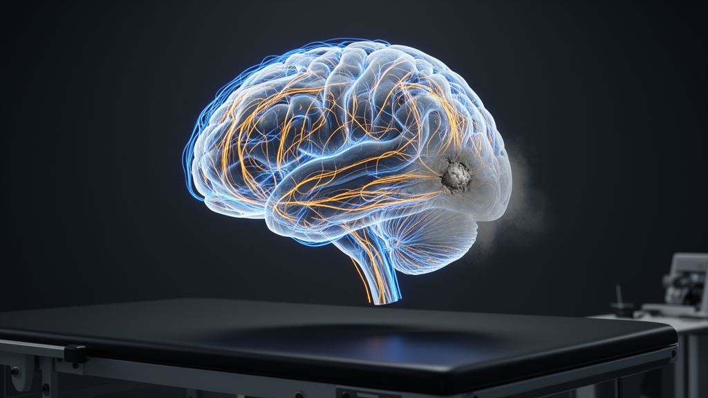
Stroke strikes roughly 12.2 million people each year, and for an estimated 36 to 47 percent of survivors the most disabling aftermath is not paralysis but cognitive impairment—losses in memory, attention, and executive function that erode independence and burden caregivers. A new narrative review published in Discover Neuroscience argues that the most promising route to better rehabilitation for these patients may lie in the connectome: the complete map of structural and functional connections linking the brain’s regions. The review, led by Nicholas Aderinto and colleagues, synthesizes how network-level imaging data could one day guide which therapy each patient receives, while candidly cataloguing why that promise is not yet clinical reality.
The core premise is mechanistic rather than speculative branding. Stroke damages not only the cells at the infarct site but also the distributed networks those cells belong to, and recovery depends critically on how those networks reorganize. The review identifies three large-scale networks consistently tied to post-stroke cognitive deficits. The default mode network, anchored in the posterior cingulate cortex, medial prefrontal cortex, hippocampus, and angular gyrus, supports wakeful rest, self-referential thought, and autobiographical memory; its disruption correlates with impaired encoding of new information and reduced insight. The frontoparietal network, built on the dorsolateral prefrontal cortex and posterior parietal cortex, carries goal-directed attention, working memory, and cognitive flexibility. The salience network, comprising the anterior insula, dorsal anterior cingulate cortex, and amygdala, filters salient stimuli and toggles activity between the other two networks. Damage to any of these, or to their interactions in the so-called triple network, produces distinct and predictable cognitive profiles.
Lesion location mediates much of this pattern. Frontal cortical lesions disrupt executive function through the frontoparietal network; parietal lesions affect the dorsal attention network and produce spatial neglect; temporal lesions impair language and episodic memory; thalamic and other subcortical lesions disturb thalamocortical circuits; and brainstem lesions interrupt arousal and cholinergic signaling with widespread attentional consequences. The review also emphasizes diaschisis—the dysfunction of structurally intact, remotely connected tissue that has lost its primary input. Remote cortical regions become hypometabolic and show neurovascular uncoupling, and the reversal of diaschisis is among the earliest contributors to spontaneous recovery, opening a time window during which rehabilitation is thought to be most effective.
From this biology the authors derive two mechanistically distinct treatment strategies. Network restoration aims to reactivate damaged circuits themselves—for example, excitatory transcranial magnetic stimulation or anodal transcranial direct current stimulation applied to the ipsilesional hemisphere to re-establish pre-injury neural activity. Network compensation instead recruits intact circuits and alternative pathways to take over lost functions, such as engaging the undamaged hemisphere or parietal networks to support language after left-hemisphere stroke. The review reports evidence that these mechanisms are neurobiologically distinct, require different timing relative to stroke onset, and likely suit different patients: those with relatively preserved ipsilesional connectivity may benefit from restoration, while severely disconnected patients may respond better to compensatory approaches. Crucially, the authors stress that this selection principle has not yet been operationalized in clinical practice; it should instead inform the design of future trials.
Where does the supporting evidence stand? For non-invasive brain stimulation, the review describes convergent findings across motor and cognitive applications: network-targeted stimulation produces more sustained neurophysiological effects than anatomically standardized protocols, and stimulation delivered during or immediately after task performance outperforms stimulation at rest, suggesting synergy with behavioral training. In multiple trials of high-frequency TMS over left dorsolateral prefrontal and inferior frontal regions, improvements in working memory, attention, and post-stroke aphasia were observed, and when fMRI connectivity mapping was used to select targets, effect sizes reportedly rose to roughly 1.5 to 2 times those of standardized targeting—a pattern the authors say has been reproduced by four independent laboratories. For tDCS, effects on cognition are small and heterogeneous, likely because individual skull and cortical anatomy scatter the delivered current; connectome-based individualization of electrode placement reportedly reduced inter-individual variance in effect sizes by about 30 to 40 percent. A fundamental limitation remains: current devices stimulate nodes, not connectivity changes in chosen directions or magnitudes.
Behavioral rehabilitation, often overshadowed by technology-driven approaches, offers some of the strongest prognostic evidence. Falconer and colleagues showed that functional connectivity within the language network predicted responsiveness to behavioral language therapy more accurately than lesion volume alone. Wilmskoetter and colleagues linked the structural integrity of the superior longitudinal and arcuate fasciculi to language recovery after intensive behavioral treatment. Zhu and colleagues, using resting-state fMRI with graph-theoretical analysis, found that disruption of hub connectivity within the dual-stream language network—not global connectivity loss—tracked aphasia severity, and that connectivity between right-hemisphere homologues and residual left dorsal language hubs predicted clinical language performance. Yet the review is explicit that these findings support prognosis, not prescription: connectomics currently identifies who is likely to respond, not which specific therapy to choose, and prospective validation is needed before connectome-guided treatment selection enters routine care.
The technological toolbox is expanding but immature. Resting-state fMRI, the workhorse of functional connectivity research, correlates network disruption with cognitive outcomes but cannot establish causality, since it measures correlations rather than direct connections. Diffusion tensor imaging traces white matter tracts and quantifies integrity through fractional anisotropy, but fails at tract crossings and is insensitive to small lesions amid post-stroke edema. Magnetoencephalography resolves network oscillations at millisecond timescale but with lower spatial resolution and specialized facility requirements. Neurofeedback and brain-computer interfaces show consistent closed-loop specificity—connectivity changes appear only when neural activity directly drives feedback—and training effects are selective to task-relevant circuits, but cognitive outcome evidence remains preliminary, with adequately powered multicenter randomized trials still lacking. Virtual reality has Cochrane-level support in motor rehabilitation but not yet for visuospatial attention or executive function.
Predictive modeling is where ambition most clearly outruns validation. Machine-learning connectome-based predictive models can estimate functional recovery at three months after stroke from connectivity data acquired at onset or one month later, with moderate group-level performance, but most models are built and validated on single-center datasets with modest samples, inconsistent reproducibility, and scanner and preprocessing variability that undermines cross-site generalizability. The review states plainly that current models are insufficiently reliable for individualized clinical decision-making. Pharmacological adjuncts fare no better: minocycline combined with stimulation showed promise only in small non-randomized trials with surrogate endpoints; observational links between low vitamin D and worse stroke outcomes have not translated into adequately powered randomized trials showing cognitive benefit; and colivelin’s evidence is entirely pre-clinical, making its application speculative.
The structural weaknesses of the field are catalogued with unusual candor. Typical connectomics rehabilitation studies enroll between 10 and 50 patients at a single center, leaving results vulnerable to publication bias. Imaging acquisition parameters—field strength, scan length, head motion, smoothing, parcellation atlases—vary widely, making meta-analysis and multi-site predictive models difficult. Reporting standards such as COBIDAS MECO exist but are incompletely adopted. Follow-up periods are usually too short to capture neuroplasticity that unfolds over months to years, and causal links between connectivity change and cognitive improvement are hard to confirm because both may reflect spontaneous recovery or compliance. The literature is also concentrated in high-income settings with 3T MRI access, leaving little direct applicability to low- and middle-income countries where stroke burden is highest.
The authors’ conclusion is measured: connectomics is not itself an intervention but a scientific basis for making interventions more efficient. Resting-state fMRI and diffusion imaging can already aid patient stratification and recovery prediction, with the strongest early findings in post-stroke aphasia. What the field needs, the review argues, are multi-center trials with standardized imaging and rehabilitation protocols, serial neuroimaging to characterize connectivity trajectories from acute through chronic phases, integration of genomic and other omics data capturing molecular determinants of plasticity, connectivity-based outcome measures embedded in behavioral trials, and resource-appropriate tools such as EEG-based connectivity measures or normative connectome databases for lower-income settings. Regulatory and ethical frameworks for AI-based decision support—covering model transparency, algorithmic bias, consent, and liability—must be developed before predictive models approach the clinic. Until then, personalized, wiring-guided cognitive rehabilitation remains a well-mapped hypothesis awaiting its validating trials.
Subject of Research: Connectome-guided approaches to personalizing cognitive rehabilitation after stroke
Article Title: Connectome guided approaches to cognitive rehabilitation after stroke
Article References: Aderinto, N., Abraham, I. C., Emmanuel, A. B., Aboje, J. E., Agbo, C. E., Okunowo, P., Bolanle, O., Adejumo, F. A., Donaldy, W., Babawale, E. A., Ajayi, A., Akata, N. M., Sule, O., Gaur, A., Kokori, E., Olatunji, G., & Aljamea, F. H. (2026). Connectome guided approaches to cognitive rehabilitation after stroke. Discover Neuroscience, 21(1), Article 100. https://doi.org/10.1186/s13064-026-00332-y
Image Credits: AI Generated
DOI: 10.1186/s13064-026-00332-y
Keywords: stroke, connectome, cognitive rehabilitation, post-stroke cognitive impairment, neuroimaging, brain stimulation, neurofeedback, machine learning, aphasia, neuroplasticity, default mode network, rehabilitation
News Source: Cassandra Pierce. (October 29, 2026). Brain Wiring Maps Could Steer Personalized Cognitive Rehab After Stroke. Scienmag.



