


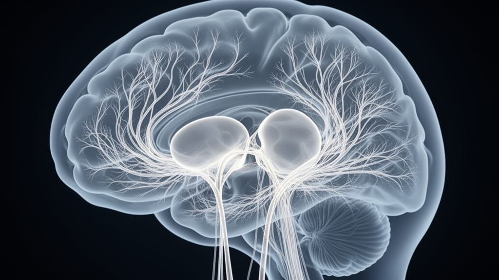
Patients with idiopathic normal pressure hydrocephalus (iNPH) show measurable alterations in the organization of their brain’s structural wiring, according to a study published in Fluids and Barriers of the CNS that used diffusion spectrum imaging (DSI) and graph-theoretical analysis to map the condition’s effects on large-scale white-matter networks. iNPH is a treatable dementia syndrome, yet the relationship between disturbed cerebrospinal fluid dynamics and disruption of the brain’s large-scale white-matter networks has remained incompletely understood.
The research team acquired DSI data from 23 patients with definite iNPH and 20 normal controls, reconstructing whole-brain structural connectomes to evaluate global and nodal topological properties. Network-based statistics were applied to identify altered subnetworks, and exploratory logistic regression and receiver operating characteristic (ROC) analyses assessed the apparent diagnostic performance of selected network metrics.
The analysis revealed a consistent pattern of increased network segregation alongside impaired global integration in the patient group. Compared with controls, patients showed a higher weighted clustering coefficient (p = 0.027) and higher transitivity (p < 0.01), both indicators of greater local segregation, together with a prolonged characteristic path length (p = 0.036), reflecting reduced global integration across the network. In network terms, the patients’ connectomes appeared more locally clustered while communication between distant regions became less efficient.
Network-based statistics pinpointed the damage more precisely: weakened structural connectivity was found primarily in interhemispheric frontal, parietal, and cingulate pathways. This pattern suggests that the connections linking the two hemispheres across the frontal, parietal, and cingulate regions bear a substantial share of the network disruption in iNPH, complementing the global topological changes observed in the connectome measures.
The study also explored whether network metrics could distinguish patients from controls. A model combining binary transitivity and the weighted assortativity coefficient, tested in the same cohort, yielded an apparent area under the ROC curve of 0.867, with sensitivity of 0.955 and specificity of 0.750. The authors emphasize that this was an exploratory, same-cohort analysis, and that the diagnostic utility of DSI-derived network metrics requires validation in larger independent cohorts before any clinical application. Overall, the findings characterize iNPH-related structural connectome alterations marked by increased segregation, impaired integration, hub reorganization, and disrupted interhemispheric connectivity, and suggest that DSI-derived network measures may serve as complementary exploratory markers of the network disruption associated with the syndrome.
Subject of Research: Structural connectome alterations in idiopathic normal pressure hydrocephalus measured with diffusion spectrum imaging
Article Title: Altered brain network properties in patients with idiopathic normal pressure hydrocephalus: a diffusion spectrum imaging study
Article References: Lv, M., Wu, Z., Du, M., Que, Y., Xu, M., Huang, H., Yu, C., Yang, X., Zhu, C., Lu, T., & Xia, J. (2026). Altered brain network properties in patients with idiopathic normal pressure hydrocephalus: a diffusion spectrum imaging study. Fluids and Barriers of the CNS. https://doi.org/10.1186/s12987-026-00870-x
Image Credits: AI Generated
DOI: 10.1186/s12987-026-00870-x
Keywords: idiopathic normal pressure hydrocephalus, diffusion spectrum imaging, structural connectome, graph theory, network-based statistics, white matter, dementia, brain networks, interhemispheric connectivity, diagnostic markers, Altered, brain
News Source: Cassandra Pierce. (October 30, 2026). Brain Wiring Disrupted in Idiopathic Normal Pressure Hydrocephalus, Imaging Study Finds. Scienmag.



