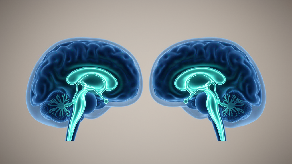
A widely used MRI measure that researchers treat as a window into the brain’s waste-clearance fluid system may be shaped far more by simple brain anatomy than by the physiology it is supposed to capture. In a cross-sectional study of 784 women drawn from the Human Connectome Project Aging and Aging Adult Brain Connectome studies, a team led by Akram Zamani and David K. Wright found that the size of the brain’s lateral ventricles was strongly and inversely associated with the diffusion tensor image analysis along the perivascular space, or DTI-ALPS, index. When ventricular volume was taken into account, apparent links between reproductive hormones and the index largely disappeared.
The DTI-ALPS index has become popular because it offers an indirect, non-invasive readout of perivascular fluid movement, the process often described as glymphatic transport. Because the measurement is taken in periventricular white matter, the authors reasoned that enlargement of the ventricles, a common feature of ageing brains, could alter the index independently of, or alongside, genuine changes in brain-fluid physiology.
To test this, the researchers used baseline data from female participants and examined whether circulating levels of three hormones, estradiol, follicle-stimulating hormone and luteinising hormone, were associated with the DTI-ALPS index. Reproductive ageing provided a natural test case, because the hormonal shifts of menopause occur alongside age-related ventricular enlargement, making it possible to ask whether hormone signals in the index are genuine or merely reflect underlying anatomy.
The analysis was built around four sequential linear regression models: an unadjusted model, a model adjusted for age, a model adjusted for age plus lateral ventricular volume, and a fully adjusted model that also included body mass index, study site and hormone replacement therapy status. Secondary analyses explored possible effects of sleep quality, APOE ε4 carrier status and choroid plexus volume. Lateral ventricular volume was derived from FreeSurfer segmentations of the structural scans.
In the unadjusted models, the hormone associations looked striking. Higher estradiol was linked to a higher ALPS index, with an estimated coefficient of 0.044 (95% CI 0.033 to 0.056), while higher follicle-stimulating hormone and luteinising hormone were each linked to lower values, with coefficients of −0.046 (95% CI −0.058 to −0.033) and −0.046 (95% CI −0.063 to −0.029) respectively. On its face, that pattern would be easy to read as evidence that the hormonal environment of midlife shapes fluid transport in the brain.
That reading did not survive statistical scrutiny. Once age was added to the models, the hormone associations changed, and after lateral ventricular volume was included they were no longer evident. Ventricular volume itself was inversely associated with the index, with a correlation coefficient of −0.71 (95% CI −0.74 to −0.67), a remarkably strong relationship for a brain-imaging measure. Including ventricular volume attenuated the estradiol effect estimates by 94% to 100% across multiple ways of parameterising the variable, and similar attenuation was seen for follicle-stimulating hormone and luteinising hormone.
The authors conclude that lateral ventricular volume was strongly associated with DTI-ALPS and statistically accounted for most of the endocrine-related variation in the index. In other words, much of what looks like hormonal influence on this fluid-transport marker may instead reflect the geometry of the ventricles next to which the measurement is taken, or biology that ventricular size and hormones share through ageing.
Importantly, the study is cross-sectional and cannot establish whether the anatomical dependence reflects measurement geometry, shared age-related biology, or both. The participants were women with a mean age of 60.0 years (SD 15.1), so the findings speak specifically to this cohort and design, and the authors note that inferences about actual brain-fluid transport from the index require independent validation. The paper, published open access in Fluids and Barriers of the CNS on 27 October 2026, was shared early as a peer-reviewed, accepted version of record in progress.
The practical message for the field is straightforward: ventricular morphology should be considered whenever the DTI-ALPS index is interpreted, and studies that report hormone or disease associations with the index without accounting for ventricular size may be capturing anatomy rather than physiology. As the metric continues to spread through the ageing and neurodegeneration literature, this analysis suggests that the ventricles sitting beside the measurement window deserve a central place in the statistical models.
Subject of Research: Dependence of the DTI-ALPS MRI index on lateral ventricular volume in aging women
Article Title: Ventricular anatomy is a major source of DTI-ALPS variability in aging females
Article References: Zamani, A., Kirk, M., Monif, M., Kapoor, M., Wells, J., Shultz, S. R., O’Brien, T. J., & Wright, D. K. (2026). Ventricular anatomy is a major source of DTI-ALPS variability in aging females. Fluids and Barriers of the CNS. https://doi.org/10.1186/s12987-026-00899-y
Image Credits: AI Generated
DOI: 10.1186/s12987-026-00899-y
Keywords: DTI-ALPS, glymphatic system, diffusion MRI, ventricular volume, aging, estradiol, FSH, luteinising hormone, Human Connectome Project, perivascular spaces, neuroimaging, women's health
News Source: Cassandra Pierce. (October 27, 2026). Brain ventricle size drives variability in a popular MRI marker of fluid transport. Scienmag.