


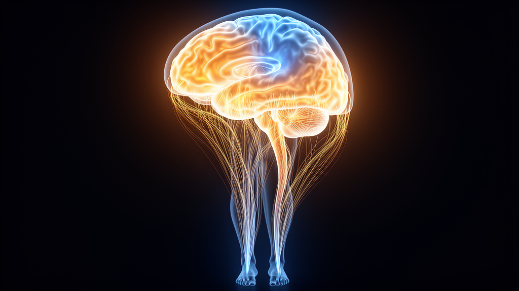
A new study published in BMC Neurology examined simultaneous electroencephalogram and electromyography recordings from Parkinson’s patients who experience freezing, tracking how cortical activity and muscle output change together across the moments before, during, and after a freezing episode.
Among three microstates identified in the walking recordings, one characterized by a hemispheric gradient showed significant modulation across gait phases. Its coverage, the proportion of time it dominated the EEG, dropped significantly during freezing compared with the pre-freezing phase.
Muscle activity followed a strikingly similar trajectory. The root mean square amplitude of the EMG signal, a measure of overall muscle activation, reached its lowest level during freezing episodes and was significantly reduced relative to both baseline walking and the pre-freezing phase. This parallel timing suggested that cortical state dynamics and motor output might be linked during these episodes.
Statistical modeling supported that link. Using linear mixed-effects modeling, the team found a significant negative association between coverage of the hemispheric-gradient microstate and EMG amplitude, pointing to a measurable coupling between cortical state and muscle output around freezing events.
The study also tested whether muscle activity alone could warn of an impending freeze. Features derived from EMG root mean square signals were used to train random under-sampling boosting classifiers, evaluated with leave-one-subject-out cross-validation. The classifiers achieved areas under the receiver operating characteristic curve of 0.62, 0.63, 0.65, 0.68, 0.77, and 0.90 at prediction horizons of 5, 4, 3, 2, 1, and 0.5 seconds before onset, respectively, with predicted freezing risk rising progressively as onset approached.
The authors conclude that the hemispheric-gradient microstate shows freezing-related modulation and an association with muscle activity, suggesting a potential bridge between cortical state dynamics and motor output in Parkinson’s disease. They also note that the EMG features showed potential for short-horizon prediction of freezing onset, though the results come from offline analyses rather than real-time, deployed systems.
Because the study analyzed recordings from Parkinson’s patients with freezing of gait, its findings describe associations within that population and do not establish causation. The modest prediction performance at longer horizons also reflects the difficulty of anticipating these episodes from muscle signals alone. Still, the converging evidence from EEG microstates and EMG offers a framework for future work on detecting and understanding freezing episodes in this disabling symptom of Parkinson’s disease.
Subject of Research: EEG microstate dynamics and EMG coupling during freezing of gait in Parkinson's disease
Article Title: EEG microstate-EMG associations during freezing of gait in Parkinson’s disease
Article References: Li, J. (2026). EEG microstate-EMG associations during freezing of gait in Parkinson’s disease. BMC Neurology. https://doi.org/10.1186/s12883-026-05587-w
Image Credits: AI Generated
DOI: 10.1186/s12883-026-05587-w
Keywords: Parkinson's disease, freezing of gait, EEG microstates, electromyography, brain-machine coupling, gait prediction, motor symptoms, machine learning, BMC Neurology, microstate-EMG, associations, during
News Source: Cassandra Pierce. (October 29, 2026). Brain Signal Patterns Shift as Freezing of Gait Strikes Parkinson’s Patients. Scienmag.



