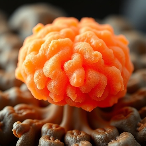
Scientists have kept human brain organoids alive and developing for more than five years, creating the longest-running laboratory model yet of the human brain’s early life. The three-dimensional tissues continued to mature in ways that closely tracked known patterns of human neurodevelopment, including changes in cell types, gene activity, epigenetic markings and neuronal communication. The findings, published in Nature, suggest that brain organoids are not merely static clusters of living cells. Under carefully engineered conditions, they can preserve a developmental “clock” and reproduce aspects of the human brain’s progression long after most laboratory models would have stopped changing. The achievement could transform how researchers study autism, schizophrenia, neurodegeneration and other disorders whose origins unfold over years rather than weeks.
The human brain develops over an unusually extended period. Although the basic neural architecture begins forming before birth, the production, specialization and communication of brain cells continue through childhood, adolescence and, in some respects, into early adulthood. Researchers have traditionally investigated this process using donated human tissue, which offers valuable but isolated snapshots, and animal models, whose brains differ from ours in anatomy, cell composition and developmental timing. Brain organoids provide a different approach. They are grown from human stem cells and self-organize into miniature, three-dimensional tissues containing multiple neural and supporting cell types. Because they can be observed repeatedly in the laboratory, organoids offer an opportunity to follow developmental change directly. Until now, however, most studies have concentrated on the earliest stages because maintaining the tissue for years has been technically difficult.
The international research team, working at Harvard University in the laboratory of Paola Arlotta, focused on the central problem of long-term survival: how to keep neurons healthy and active without forcing the tissue into an artificial developmental standstill. Standard culture systems can sustain organoids for considerable periods, but prolonged growth often leads to declining neuronal activity and imbalances in the cell populations. The researchers altered the composition of the culture medium to better support spontaneous electrical activity, a characteristic feature of developing human neurons. Neurons in the human brain do not wait for sensory experiences before becoming active; they generate internally coordinated patterns that help shape connections. By preserving this activity, and by maintaining the organoids in an isolated environment with continuous specialist care, the team extended the functional life of the tissues beyond five years.
The organoids were not simply stored in a living state. At selected time points, the researchers used a range of molecular and cellular measurements to determine whether the cultures were continuing to develop. They identified the cell types present, measured patterns of gene expression, examined epigenetic modifications and recorded neuronal activity. Gene-expression analysis revealed that neural populations emerged in an order resembling the sequence observed in human brain development. Early neural progenitors gave rise to increasingly specialized neurons, followed by the appearance and expansion of glial populations. At the same time, neurons developed more elaborate connections, indicating that the tissue was becoming more structurally and functionally complex rather than merely surviving through slow biological maintenance.
One of the most compelling signs of continued maturation came from epigenetics, the system of chemical modifications that regulates how DNA is used without changing the genetic code itself. Among these modifications are changes to DNA methylation, which can accumulate according to developmental age and help determine which genes are active or silent. In human brain tissue, these patterns shift in a relatively predictable sequence as cells mature. The long-lived organoids displayed comparable changes over time. After approximately one year, the cultures began showing molecular features associated with stages that normally occur after birth. Their cells therefore appeared to retain an internal record of developmental time, even though they were growing outside the body and without the full range of signals supplied by a developing human organism.
The researchers also tested whether this developmental history remained embedded in mature cells. They separated cells from older organoids and allowed them to grow again under conditions that encouraged renewed organization. Cells from the older tissues continued to generate populations associated with later developmental stages. When the investigators combined older cells with younger cells, however, the older cells regained some capacity to produce neurons, although the neurons belonged predominantly to later stages of the developmental sequence. The result suggests that cellular potential is not erased as the organoid ages. Instead, cells retain a molecular memory of where they are in developmental time, while environmental signals can influence how that potential is expressed.
This finding could offer a new way to investigate the boundary between neurodevelopment and repair. In the human brain, the production of new neurons is generally most active early in life and declines sharply in many regions as the brain matures. Later development increasingly favors the production of glial cells, which support neurons, regulate the cellular environment and contribute to immune defense and repair. If scientists can identify the signals that allow older organoid cells to resume neuron production, they may gain clues about how regenerative programs are switched off in the human brain—and whether some of them can be reactivated. Such work could eventually inform studies of neurodegenerative diseases, although the organoids remain simplified models and cannot yet reproduce the full complexity of a living brain.
The extended lifespan also makes the organoids potentially valuable for studying disorders that cannot be captured by examining only early development. Autism spectrum disorder and schizophrenia, for example, involve complex interactions among genetic risk, cellular maturation and neural circuitry. Some disease-related differences may not become visible until particular cell types emerge or networks begin communicating over longer periods. Long-lived organoids derived from different genetic backgrounds could allow researchers to compare developmental trajectories, identify when abnormalities first appear and test whether candidate drugs correct specific cellular or electrical defects. Their reproducibility is especially important: reliable cultures could make it easier to distinguish genuine disease-related effects from differences caused by experimental variation.
The models still have significant limitations. They lack a full blood supply, and their internal organization does not reproduce the layered architecture of the human cerebral cortex with complete accuracy. They also do not receive the sensory input, hormonal signals, immune interactions and body-wide feedback that influence brain development in a living person. The researchers are now exploring whether environmental stimulation, including carefully controlled exposure to light, can promote further maturation and more organized neural activity. Improving vascularization and cortical layering will be essential if organoids are to model later developmental stages with greater fidelity. Even so, the new work demonstrates that human neural tissue grown in the laboratory can remain biologically dynamic for years, opening a much longer window for observation than previously thought possible.
The broader significance lies in the organoids’ ability to record the passage of developmental time. Their changing cell populations, gene-expression programs, epigenetic signatures and electrical behavior together form a biological timeline that resembles the sequence of human brain maturation. Researchers now have a platform for asking not only how brain cells are made, but also how they remember when they were made and how their surroundings influence what they become. “We now know that these models have the capacity to continue developing for years,” says Noelia Antón-Bolaños, an assistant professor at University Medical Center Utrecht and one of the study’s leaders. The next challenge is to provide conditions that allow that capacity to unfold more completely, bringing laboratory models closer to the developmental reality of the human brain.
Subject of Research: Lab-produced tissue samples
Article Title: Human brain organoids record the passage of time over multiple years
News Publication Date: 19-Aug-2026
Web References: https://doi.org/10.1038/s41586-026-10877-x; https://research.umcutrecht.nl/researchers/noelia-anton-bolanos/
References: Nature, “Human brain organoids record the passage of time over multiple years,” DOI: 10.1038/s41586-026-10877-x
Image Credits: D.C.N. van der Heijden / UMC Utrecht
Keywords: brain organoids, human brain development, stem cells, neurodevelopment, epigenetics, neuronal activity, autism, schizophrenia, neurodegenerative disease, regenerative neuroscience
Tags: advancing neuroscience with long-term organoid studiesBrain organoids longevitydevelopmental timing in brain organoidsepigenetic changes in brain developmenthuman neurodevelopment modelinglaboratory simulation of early human brain developmentlong-term brain tissue culturemodeling neurodegenerative diseases with brain organoidsneural cell maturation in organoidsneuronal communication in lab-grown brainsorganoid-based autism and schizophrenia researchstem cell-derived human brain models