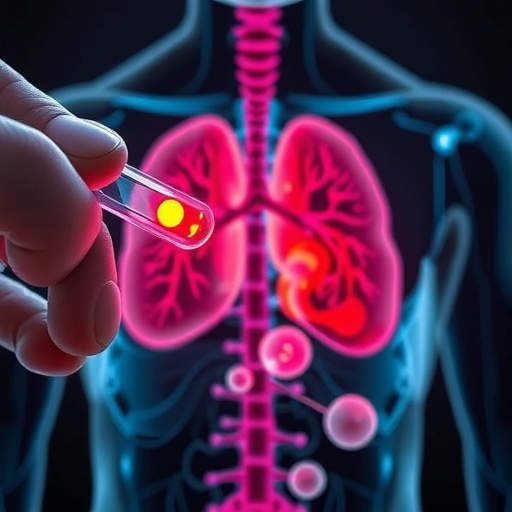
Artificial intelligence can estimate the biological age of individual human organs by reading microscopic patterns in tissue architecture, according to a large study led by researchers at the CeMM Research Center for Molecular Medicine of the Austrian Academy of Sciences and the Ludwig Boltzmann Institute for Network Medicine at the University of Vienna. The research, published in Nature Medicine, used more than 25,000 histological images from 983 people and examined 40 tissue types, ranging from the brain and heart to the lung, kidney, pancreas, skin, intestine, and reproductive organs. The resulting “tissue clocks” predicted biological age with a mean error of 4.9 years, offering a new way to study how aging unfolds inside the body. Unlike conventional measures based primarily on DNA methylation or gene expression, these models analyze the physical organization of cells and structures visible in tissue sections. The findings suggest that organs do not follow a single aging timetable and that disease-related changes may be detectable through tissue architecture long before they become obvious through routine clinical assessment.
The study addresses a fundamental problem in aging research: chronological age is an imperfect measure of biological condition. Two people with the same number of years may have organs with very different levels of cellular stress, structural deterioration, and disease burden. To investigate this gap, the researchers turned to the Genotype-Tissue Expression Project, or GTEx, a large resource containing molecular and tissue data collected from human donors. The investigators converted tissue specimens into high-resolution digital images and divided them into approximately 480 million individual image tiles for computational analysis. Each tile contained localized information about features such as cell density, tissue organization, nuclei, blood vessels, connective tissue, and microscopic lesions. State-of-the-art computer vision models then learned to associate these patterns with the donor’s chronological age. Importantly, the models were not simply trained to recognize one obvious visual marker of aging. Instead, they identified complex combinations of spatial features distributed throughout each tissue type.
Age emerged as the strongest factor shaping tissue appearance across all 40 tissues analyzed. This result indicates that histological architecture contains a persistent record of the aging process, even when the changes are too subtle or diffuse for a pathologist to interpret consistently by eye. In biological terms, the models appear to capture the cumulative consequences of processes such as altered cellular composition, extracellular matrix remodeling, chronic inflammation, fibrosis, vascular change, and declining tissue repair. The tissue clocks translated those visual patterns into an estimate of biological age for a specific organ. A liver-derived prediction, for example, was intended to describe the aging state of the liver rather than provide a generalized age estimate for the entire person. This organ-specific design allowed the researchers to compare aging trajectories across tissues and to identify cases in which one organ appeared substantially older or younger than expected from chronological age.
The resulting trajectories revealed that aging is neither uniform nor linear throughout the body. The lung, kidney, pancreas, and adrenal gland showed evidence of accelerated structural change between approximately 20 and 40 years of age, suggesting that some organs may undergo important biological transitions relatively early in adulthood. Other tissues followed more complicated patterns, with periods of accelerated aging emerging later in life or appearing in several distinct phases. The uterus displayed a particularly pronounced shift around menopause, a finding consistent with major hormonal and physiological changes that influence tissue structure. These results challenge the idea that aging can be represented by one universal curve applying equally to every organ. Instead, each tissue appears to combine systemic influences, such as inflammation and metabolic status, with local factors including workload, exposure to injury, regenerative capacity, and hormonal regulation.
The clocks also captured relationships between tissue age and established markers of biological aging. Older predicted tissue age was associated with telomere shortening, greater histopathological abnormalities, and a higher number of chronic diseases. Telomeres are protective DNA structures that generally become shorter as cells divide and experience cumulative stress, although their relationship with aging is complex and varies across tissues. The connection between histological age and telomere length suggests that visual tissue architecture may integrate several molecular processes into a single measurable phenotype. The models also detected structural signatures associated with specific clinical conditions. Kidney failure, for instance, was linked to accelerated aging signals across multiple tissues, indicating that severe organ dysfunction may have effects beyond the affected organ itself. Diabetes produced especially pronounced changes in the pancreas, where long-term metabolic stress can influence endocrine cells, blood vessels, and surrounding tissue organization.
Because tissue sampling is invasive and cannot be performed routinely for every organ, the researchers next investigated whether tissue-specific aging information could be inferred from blood. They linked blood gene-expression profiles to the histologically derived tissue-age gaps of the same individuals. A tissue-age gap represents the difference between an organ’s predicted biological age and the person’s chronological age. Using these paired data, the team developed models that attempted to predict organ-specific age gaps from a routine blood sample alone. This approach does not mean that blood directly contains a microscopic image of the brain, kidney, or pancreas. Rather, circulating blood cells and molecules reflect systemic signals generated by tissues throughout the body. Changes in immune activity, metabolism, stress responses, and tissue injury can alter gene-expression patterns in blood, creating an indirect molecular readout of organ condition.
The blood-based predictors identified disease-associated aging patterns in several conditions, including Alzheimer’s disease, Crohn’s disease, cystic fibrosis, vasculitis, diabetes, and stroke. In Alzheimer’s disease, the strongest signal was concentrated in the brain-specific model, whereas Crohn’s disease was associated with accelerated aging patterns across the gastrointestinal tract. These findings suggest that different diseases leave distinct combinations of systemic and tissue-linked signals in circulating blood. If validated in larger and more diverse populations, such predictors could eventually support minimally invasive monitoring of disease progression or treatment response. A blood test based on this principle might not replace imaging, biopsy, or clinical examination, but could help identify patients who require closer evaluation. It could also provide a way to track changes over time when repeated tissue sampling would be impractical or unsafe.
The technical advance lies in connecting three layers of biological information: histological architecture, molecular activity, and clinical phenotype. Histology shows what cells and tissues look like; gene expression indicates which biological programs are active; and clinical data records disease, treatment, and health outcomes. By training models across these layers, the researchers created a framework in which visual patterns learned from tissue can be translated into blood-based measurements. Deep-learning systems are particularly suited to this task because they can analyze subtle spatial relationships that conventional image-processing methods may overlook. However, the models remain statistical tools rather than direct measurements of aging mechanisms. Their predictions may be influenced by differences in tissue handling, sampling location, image quality, donor demographics, and the medical circumstances surrounding tissue collection. External validation will therefore be essential before the clocks can be used in routine diagnosis.
The researchers emphasize that the tissue clocks are most valuable not as a simple score of whether someone is “old” or “young,” but as instruments for mapping how aging differs from organ to organ. Individuals whose tissues show structural changes substantially ahead of their chronological age could represent important biological outliers. Studying these outliers may reveal why some people remain resilient despite chronic stress while others develop disease earlier in life. Future research will need to test whether tissue-age gaps predict future illness, respond to lifestyle changes or therapies, and remain reliable across populations that were not fully represented in the GTEx resource. The models will also need careful calibration to distinguish normal variation from clinically meaningful pathology. Even with those limitations, the study provides a powerful new view of aging: not as a single process advancing at the same speed everywhere, but as a network of organ-specific transformations that can be read from tissue images and, potentially, monitored through blood.
Subject of Research: Human tissue samples
Article Title: “Histological aging signatures for monitoring tissue-specific aging and disease”
News Publication Date: 14 August 2026
Web References: https://doi.org/10.1038/s41591-026-04566-5
References: Genotype-Tissue Expression (GTEx) Project; Nature Medicine, DOI: 10.1038/s41591-026-04566-5
Keywords: Histological analysis; Tissue samples; Computational biology; Artificial intelligence; Biological aging; Tissue clocks; Digital pathology; Organ aging; Blood biomarkers; Machine learning
Tags: advanced tissue imaging for age estimationaging prediction in multiple organsAI-driven health diagnosticsbiological age estimation from histologyearly detection of disease-related tissue changesmicroscopic tissue pattern analysisnon-invasive organ health monitoringorgan aging prediction using artificial intelligenceorgan-specific aging markerstissue architecture analysis for agingtissue clocks for aging assessmenttissue structure and aging correlation