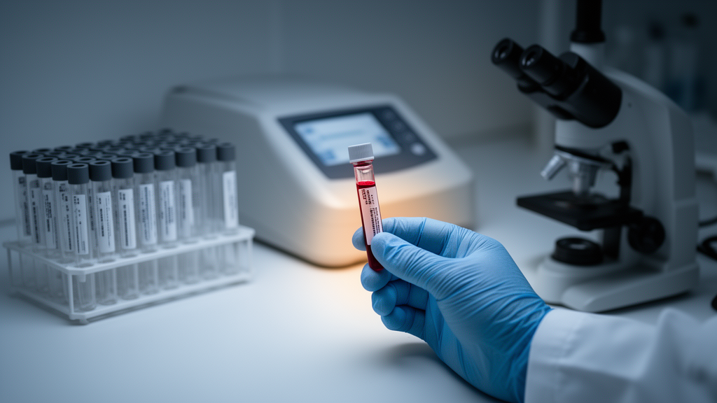
A simple blood test measuring two neural proteins together with a patient’s age could help doctors distinguish low-grade from high-grade gliomas before surgery, according to a new cross-sectional study published in the Journal of Translational Medicine.
Gliomas are the most prevalent primary malignant tumors of the central nervous system in adults, and accurate grading is crucial for selecting targeted treatment strategies and predicting disease progression. The study enrolled 89 glioma patients, 70 patients with brain metastases, and 81 healthy controls, quantifying serum levels of four neural tissue markers—GFAP, S100B, NfL, and UCH-L1—using chemiluminescence immunoassay.
The results showed that GFAP, S100B, and UCH-L1 were significantly elevated in glioma patients compared with both brain metastases patients and healthy controls, with all comparisons reaching p < 0.001. NfL was elevated in glioma patients versus healthy controls (p < 0.0001) but showed no significant difference between glioma and brain metastases patients (p > 0.05), limiting its usefulness for that specific differential diagnosis.
For detecting glioma against healthy controls, S100B and UCH-L1 achieved good discrimination, with areas under the receiver operating characteristic curve of 0.894 and 0.856, respectively. However, the researchers reported that these two markers demonstrated limited utility when the task was to differentiate glioma from brain metastases.
GFAP showed a moderate positive correlation with tumor volume (r = 0.54, 95% CI: 0.380–0.676, p < 0.001), suggesting that serum levels of this glial protein track with the amount of tumor tissue present. Levels of GFAP, NfL, and UCH-L1 were also associated with WHO grade and IDH mutation status, two key molecular and histological features that guide glioma management.
The central finding of the study came from multivariate logistic regression, which identified GFAP, NfL, and age as independent predictors of high-grade glioma. The integrated GFAP-NfL-age model achieved an AUC of 0.916 (95% CI: 0.859–0.973, p < 0.001) for distinguishing low-grade from high-grade gliomas, indicating strong discriminatory performance in this cohort.
Because models fitted to a single dataset can overestimate performance, the researchers applied bootstrap validation. The optimism-corrected AUC was 0.893 (95% CI: 0.827–0.962), only modestly below the apparent value, which supports the internal robustness of the model while leaving external validation in independent cohorts as a necessary next step.
The authors conclude that serum GFAP and NfL, combined with age, provide promising non-invasive diagnostic accuracy for preoperative glioma grading. As a cross-sectional study, the findings establish associations within the enrolled population rather than proving clinical benefit, and the model’s performance will need confirmation in prospective settings before such a blood-based approach could inform routine diagnostic decisions.
Subject of Research: Non-invasive glioma grading using serum biomarkers GFAP and NfL
Article Title: Serum GFAP and NfL for non-invasive differentiation of low-grade and high-grade gliomas
Article References: Zhuang, T., Yu, P., Dong, J., Xie, Y., Yue, X., Wei, Z., Tao, Z., & Wang, X. (2026). Serum GFAP and NfL for non-invasive differentiation of low-grade and high-grade gliomas. Journal of Translational Medicine. https://doi.org/10.1186/s12967-026-09135-5
Image Credits: AI Generated
DOI: 10.1186/s12967-026-09135-5
Keywords: glioma, GFAP, neurofilament light, biomarkers, brain tumors, glioma grading, liquid biopsy, diagnostic accuracy, IDH mutation, Journal of Translational Medicine, Serum, non-invasive
News Source: Ophelia Keating. (October 27, 2026). Blood Test Combining GFAP, NfL and Age Shows Promise for Grading Gliomas Before Surgery. Scienmag.