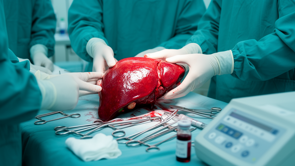
When a donor liver is finally reconnected to a recipient’s blood supply, the operating room holds its breath. Blood floods back into an organ that has spent hours chilled and starved of oxygen, and the sudden return triggers ischemia-reperfusion injury, a burst of oxidative and inflammatory damage that can determine whether the new liver thrives or falters. Now a team at the University of California, San Francisco has tracked a blood protein through that perilous moment and found that how fast it rises, rather than how high it ultimately climbs, may reveal which patients are headed for early allograft dysfunction, one of the most feared complications of liver transplantation.
The protein, pentraxin-3, is a long pentraxin, a humoral pattern recognition molecule produced not by the liver like its famous cousin C-reactive protein, but by endothelial cells and leukocytes. That origin matters. Pentraxin-3 sits at the intersection of three endothelial functions that go haywire after reperfusion: it mediates innate immune responses, modulates P-selectin-dependent leukocyte recruitment to the vessel wall, and influences coagulation. Because early allograft dysfunction is thought to arise downstream of endothelial inflammation and dysregulated coagulation, the researchers hypothesized that circulating pentraxin-3 levels measured during transplant surgery would be associated with the condition.
In a retrospective cohort study published in Frontiers in Transplantation, Alexandria Page, Michael Bokoch, Dieter Adelmann, Rachel Lin, Judith Hellman and Kerstin Kolodzie quantified plasma pentraxin-3 in 76 liver transplant recipients and 7 healthy living donors recruited to a biobank between May 2021 and August 2025. Samples were drawn at four intraoperative time points: the start of surgery, the end of liver dissection, 15 minutes after reperfusion, and 60 to 120 minutes after reperfusion. Control samples from the living donors, who underwent donor hepatectomy but no reperfusion of an ischemic graft, were collected at the start and end of their operations. Pentraxin-3 was measured by enzyme-linked immunosorbent assay, and the exposure was examined both as absolute concentrations and as delta values, the change between paired time points.
The numbers tell a striking story. At the start of surgery, transplant recipients already carried roughly three times more pentraxin-3 than healthy donors, with a median of 1.72 nanograms per milliliter against 0.57 in controls, a difference that was highly significant. That baseline elevation tracks with the proinflammatory state of end-stage liver disease: initial pentraxin-3 correlated weakly but significantly with the recipients’ MELD-Na scores, a measure of pretransplant illness severity. In the 50 patients with complete serial samples, median pentraxin-3 climbed from 1.74 nanograms per milliliter at the start of surgery to 6.70 fifteen minutes after reperfusion and 15.2 at the final draw, a rise that was overwhelmingly driven by the post-reperfusion period. The increase between the two post-reperfusion draws was more than double the increase from the start of surgery to reperfusion, suggesting that the return of blood to the graft, not the surgery itself, is the dominant trigger.
The contrast with the living donors is perhaps the study’s most vivid finding. These donors underwent major hepatic surgery with substantial tissue trauma, yet their pentraxin-3 rose only modestly, from 0.57 to 1.78 nanograms per milliliter by the end of their operations. Recipients, by comparison, finished with a median of 15.9 nanograms per milliliter, nearly an order of magnitude higher. Because both groups experienced significant surgical stress but only the recipients experienced reperfusion of an ischemic graft, the authors speculate that ischemia-reperfusion injury of the liver graft is what ignites the pentraxin-3 surge. Their data are consistent with a recent transcriptional study showing that pentraxin-3 mRNA expression rises in human liver biopsies in the hours after reperfusion, and that in mouse models of hepatic ischemia-reperfusion injury, higher pentraxin-3 expression was associated with greater pathological damage.
But the most clinically consequential result concerns not the final concentration but the trajectory. Early allograft dysfunction, defined by the Olthoff criteria, occurred in 17 of the 51 patients with final samples, a rate of 33 percent that mirrors the roughly 25 percent incidence reported nationally. In patients who developed the condition, the delta-pentraxin-3 between 15 minutes and 60 to 120 minutes after reperfusion was significantly greater, with a median of 12.3 nanograms per milliliter against 6.59 in those who did not develop dysfunction. Yet the final absolute pentraxin-3 level showed no significant difference between the two groups. In other words, the speed of the rise after reperfusion, not the peak, carried the signal. The area under the receiver-operating characteristic curve was 0.67 for delta-pentraxin-3 and 0.63 for the final level, indicating modest discrimination that would need improvement before clinical use.
The mechanism the authors propose is plausible and testable. Reperfusion activates cells within the donor liver, including endothelial cells, lymphocytes and Kupffer cells, the liver’s resident macrophages, which then synthesize and release pentraxin-3. Alternatively, substances flushed from the graft during reperfusion may induce systemic production. Either way, rising pentraxin-3 is thought to promote endothelial dysfunction, which in turn contributes to ischemia-reperfusion-induced allograft injury. In vitro evidence supports this: pentraxin-3 has been shown to induce vascular endothelial dysfunction through a P-selectin and matrix metalloproteinase-1 pathway. Interventions targeting the same axis, including P-selectin-blocking agents and donor statin treatment, have shown promise in early clinical trials, suggesting that pentraxin-3 may mark a pathway that is already druggable.
A genuinely unexpected finding emerged when the researchers stratified patients by graft preservation method. Recipients of deceased donor livers that had undergone normothermic machine perfusion, a technique that pumps warm oxygenated blood through the graft before implantation, showed significantly higher delta-pentraxin-3 after reperfusion, with a median of 12.7 nanograms per milliliter, than recipients of living donor grafts, whose median was 5.79. Absolute final levels did not differ significantly. The authors speculate that machine perfusion may activate downstream inflammatory pathways earlier than static cold storage, consistent with other reports of increased endothelial activation during perfusion and with bulk RNA sequencing data showing pentraxin-3 among the top 40 differentially expressed genes in liver biopsies before and after normothermic perfusion. An alternative explanation is confounding: machine perfusion cohorts increasingly receive higher-risk grafts, including organs from donation after circulatory death, and indeed all six recipients of DCD grafts with final samples developed early allograft dysfunction.
The study has clear limitations. It was retrospective, single-center, and modest in size, with high proportions of living donor and machine-perfused transplants that precluded adjusted stratified analysis. The sequential paired sampling was limited by research staff availability during out-of-hours surgery, and the discrimination of delta-pentraxin-3 for early allograft dysfunction was low. The authors also note that pentraxin-3’s role is genuinely dual: exogenous administration or overexpression of the protein has been reported to increase survival in mice with endotoxin-induced shock, suggesting a protective function in some acute inflammatory contexts even as it appears deleterious in hepatic ischemia-reperfusion injury. No association was found between pentraxin-3 and the Model for Early Allograft Function score, possibly because that continuous measure uses inverse natural log-transformed biomarkers while the Olthoff criteria use binary thresholds, weighting biomarker components differently.
Still, the findings open a window onto a physiological event that has been difficult to observe in real time. Pentraxin-3 rises over the course of liver transplantation, peaks after reperfusion, and its post-reperfusion velocity, not its ceiling, is what distinguishes patients who go on to early graft dysfunction. If future prospective studies confirm that delta-pentraxin-3 measured in the first hours after reperfusion can identify patients at risk, clinicians may gain a biomarker that allows targeted intervention while the graft is still salvageable, whether through endothelial-protective agents, refined perfusion protocols, or personalized postoperative monitoring. For a complication that affects a quarter of all liver transplants and sits on the path from ischemia-reperfusion injury to graft loss, that would be no small advance.
Subject of Research: Pentraxin-3 dynamics during liver transplantation and association with early allograft dysfunction
Article Title: Pentraxin-3 dynamics during liver transplantation and association with early allograft dysfunction
Article References: Page, A., Bokoch, M. P., Adelmann, D., Lin, R., Hellman, J., & Kolodzie, K. (2026). Pentraxin-3 dynamics during liver transplantation and association with early allograft dysfunction. Frontiers in Transplantation, 5, Article 1920891. https://doi.org/10.3389/frtra.2026.1920891
Image Credits: AI Generated
DOI: 10.3389/frtra.2026.1920891
Keywords: Pentraxin-3, dynamics, during, liver, transplantation, association, early, allograft, dysfunction, scientific research
News Source: Drew Townsend. (October 17, 2026). Blood protein surge after liver reperfusion may flag early transplant graft failure. Scienmag.