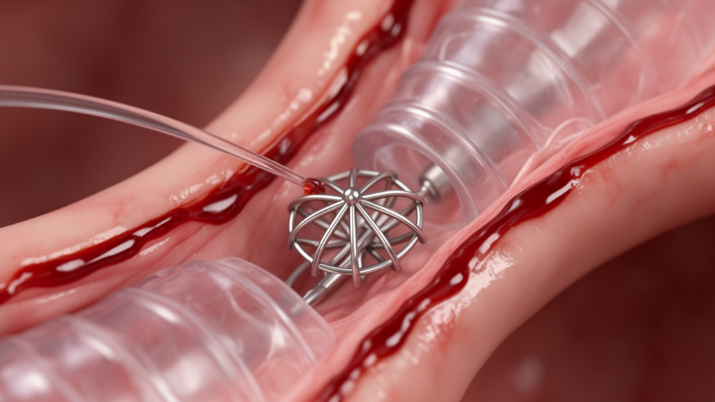
A case report demonstrates that autologous blood patch pleurodesis successfully sealed a persistent air leak following bronchoscopic lung volume reduction while allowing all implanted endobronchial valves to remain in place. The procedure, reported in Respirology Case Reports, resolved an air leak that had persisted for nine days despite active chest tube drainage, with bubbling ceasing within twenty-four hours after instillation. By avoiding the removal of endobronchial valves, the medical team successfully preserved target-lobe atelectasis and documented subsequent improvements in lung function and exercise capacity over a six-month follow-up period.
Bronchoscopic lung volume reduction using endobronchial valves is established as a minimally invasive intervention designed for patients with severe emphysema, offering measurable improvements in respiratory symptoms, functional exercise capacity, and objective lung function measurements. However, procedural pneumothorax represents a recognized adverse event, occurring in approximately 4.2 percent to 34.4 percent of patients undergoing the procedure. When a persistent air leak arises after valve placement, management typically requires prolonged chest tube drainage. When the leak remains significant, clinicians are frequently forced to extract one or more implanted valves, which facilitates pleural defect closure but risks reversing intended lobar atelectasis and sacrificing therapeutic benefit.
The patient described in the clinical report was a 60-year-old man diagnosed with Global Initiative for Chronic Obstructive Lung Disease Stage IV chronic obstructive pulmonary disease, referred due to worsening exertional dyspnoea despite receiving optimal inhaled medical therapy. His medical background included a 52.5 pack-year history of cigarette smoking, a modified Medical Research Council dyspnoea grade of III, and a baseline six-minute walk distance of 324 meters, during which his oxygen saturation dropped to 86 percent. Pre-interventional physiological assessments documented a post-bronchodilator forced expiratory volume in one second of 0.78 liters, corresponding to 24.7 percent of predicted normal values, a residual volume of 207.4 percent predicted, and a total lung capacity measuring 128.0 percent predicted.
Diagnostic chest computed tomography demonstrated severe emphysematous destruction displaying an upper-lobe predominance, while quantitative StratX fissure analysis confirmed greater than 95 percent structural completeness across both major fissures. Following physiological confirmation of absent collateral ventilation using the Chartis balloon catheter system, interventionists deployed 4.0-millimeter Zephyr endobronchial valves into the patient’s left bronchial tree, targeting the B1 plus 2, B3a, B3b plus c, B4, and B5 segmental bronchi. Initial chest radiography obtained four hours following bronchoscopic valve placement revealed a left-sided pneumothorax, prompting immediate placement of an 18-French chest tube managed under a water seal.
Because the air leak failed to resolve, clinical staff subsequently applied active negative pressure suction of minus 12 centimeters of water to the drainage system. Despite continuous tube thoracostomy drainage, the air leak persisted alongside residual pneumothorax and worsening subcutaneous emphysema visualized on repeat chest radiographs and computed tomography scans obtained on post-procedure Day 7. By Day 9, bubbling remained evident in the drainage system during expiration, meeting the diagnostic threshold for Grade 2 expiratory air leak according to the standard Cerfolio classification system.
Faced with an unresolved leak that would conventionally prompt valve removal, the clinical team elected to perform autologous blood patch pleurodesis prior to extracting any implanted devices. Although clinical practice guidelines support autologous blood patching as an option for persistent air leaks complicating secondary spontaneous pneumothorax, its role in managing post-bronchoscopic lung volume reduction leaks remains poorly defined and rarely described before valve removal. The procedure involved instilling 100 milliliters of the patient’s own peripheral blood directly through the indwelling chest tube into the pleural space.
Following blood instillation, the persistent air leak resolved completely within 24 hours, and subsequent monitoring revealed no immediate procedural adverse events. Following a successful clamp trial to ensure pleural stability, the chest tube was removed on post-procedure Day 16, with serial imaging demonstrating no pneumothorax recurrence. Follow-up chest computed tomography performed three months after the initial intervention confirmed sustained left upper lobe atelectasis alongside complete resolution of the pneumothorax. Functional re-evaluation at six months revealed that the patient’s six-minute walk distance had increased from 324 meters to 533 meters, while his forced expiratory volume in one second improved from 24.7 percent to 34.2 percent of predicted values.
Discussing the physiological mechanism, the authors noted that autologous blood patch pleurodesis provides a mechanical patch effect driven by localized fibrin clot formation over the visceral pleural defect. This mechanical adhesion presents a distinct advantage in post-volume-reduction settings, where deliberate target-lobe atelectasis prevents full parenchymal re-expansion and impedes conventional pleural apposition. The authors referenced prior observations indicating that autologous blood patching can remain clinically effective even when lung re-expansion is incomplete, although patients with deflated lungs or large residual pleural cavities may require repeated instillations beyond a single 50-to-100-milliliter instillation.
The authors contrasted their management with a previously published conference abstract describing autologous blood patching following valve-induced pneumothorax, in which two of three implanted valves were extracted on Day 7 before patching was attempted on Day 28. In contrast, performing blood instillation on Day 9 in the present case permitted complete retention of all five valves, supporting the viability of blood patching as an early valve-preserving strategy. While infectious pleuropulmonary complications represent a theoretical hazard following autologous blood instillation, no infectious sequelae were observed in this patient, though the authors emphasized that careful post-procedure surveillance remains mandatory.
The authors underscored the inherent limitations of their report, cautioning that observations derived from an isolated clinical case cannot establish comparative efficacy, determine optimal intervention timing, or define precise patient selection criteria, nor can spontaneous pleural healing be entirely excluded. Nonetheless, because the air leak had persisted through nine days of continuous chest tube drainage and resolved within a single day following blood instillation, the authors concluded that autologous blood patch pleurodesis may serve as an effective, minimally invasive intermediate strategy to preserve implanted valves and their intended therapeutic benefits.
Diagnostic evaluations before the bronchoscopic procedure underscored the severity of the patient’s respiratory compromise and hyperinflation. Initial chest radiography demonstrated marked bilateral pulmonary hyperinflation accompanied by distinct diaphragmatic flattening, consistent with severe emphysematous hyperinflation. Corresponding computed tomography imaging confirmed that emphysematous destruction was concentrated predominantly within the upper lobes. To achieve targeted lobar collapse while honoring anatomical pathways, bronchoscopists positioned individual 4.0-millimeter Zephyr endobronchial valves across five specific segmental airways: the left B1 plus 2, B3a, and B3b plus c segmental bronchi, as well as the left B4 and B5 segmental bronchi. StratX analysis had verified that fissure integrity exceeded 95 percent across both major fissures, and Chartis balloon assessments validated the complete absence of collateral ventilation prior to device deployment.
Serial radiographic examinations obtained throughout the post-intervention course documented the persistent nature of the pleural defect. Computed tomography and chest radiographs performed on post-procedure Day 7, prior to blood patch pleurodesis, highlighted the distinct margin of the incompletely re-expanded lung alongside persistent subcutaneous emphysema tracking through the soft tissues. Although surgical interventions have previously been described to address persistent post-procedural pneumothorax without removing endobronchial devices, operative management presents substantial clinical hazards for this fragile demographic. Candidates undergoing bronchoscopic volume reduction typically possess advanced emphysema and severely diminished physiological reserve, which makes invasive thoracic surgery highly unfavorable. Consequently, clinical teams require less invasive interventional alternatives that can manage continuing air leaks while avoiding the anatomical and physiological burdens associated with operative pleural repair.
International clinical practice guidelines currently recognize autologous blood patch pleurodesis as an acceptable therapeutic modality for persistent air leaks in secondary spontaneous pneumothorax, though the formal level of supporting evidence remains classified as low. The mechanism of autologous blood patching relies on a physical patch effect, wherein localized fibrin clot formation adheres directly over the visceral pleural defect. This biological sealing mechanism is especially relevant following bronchoscopic reduction, where deliberate lobar atelectasis naturally restricts complete parenchymal re-expansion and inhibits visceral and parietal pleural apposition. Previous clinical evaluations by Ando and colleagues established that autologous blood instillations can successfully control air leaks even in the absence of full lung re-expansion. However, those reports also revealed that individuals presenting with deflated lung parenchyma frequently required repeated blood administrations. As a result, the authors emphasized that a standard single instillation of 50 to 100 milliliters may prove insufficient in cases characterized by extensive lung collapse and substantial residual pleural space, necessitating careful case-by-case clinical evaluation.
Safety considerations represent another critical dimension when administering blood patches in the presence of indwelling endobronchial prostheses. Potential infectious pleuropulmonary complications remain an acknowledged clinical concern following pleural blood instillation; nevertheless, the authors noted an absolute lack of published data specifically assessing whether this infectious hazard increases when autologous blood is introduced alongside retained endobronchial valves. Although no infectious sequelae developed in this patient, rigorous clinical and radiographic monitoring remains essential whenever blood pleurodesis is attempted in the setting of permanent airway implants. The timing of the intervention also distinguishes this outcome from earlier medical literature. A previous conference abstract detailed autologous blood patching for valve-induced pneumothorax, but that attempt occurred on post-procedure Day 28 after two of the three implanted devices had already been removed on Day 7. By intervening much earlier on Day 9, the present report provides practical evidence supporting autologous blood patching as an early valve-preserving strategy before device removal is initiated.
Subject of Research: Autologous blood patch pleurodesis for persistent air leak after bronchoscopic lung volume reduction
Article Title: Autologous Blood Patch Pleurodesis Without Valve Removal for Persistent Air Leak After Bronchoscopic Lung Volume Reduction: A Case Report
Article References: Sasaki, K., Takashima, Y., Nakamura, T., Harabayashi, W., Komori, T., Yoshikawa, S., Matsumoto, M., Kimura, H., Shinagawa, N., & Konno, S. (2026). Autologous Blood Patch Pleurodesis Without Valve Removal for Persistent Air Leak After Bronchoscopic Lung Volume Reduction: A Case Report. Respirology Case Reports, 14(11), Article e70801. https://doi.org/10.1002/rcr2.70801
Image Credits: AI Generated
DOI: 10.1002/rcr2.70801
Keywords: autologous blood patch, pleurodesis, bronchoscopic lung volume reduction, endobronchial valves, persistent air leak, pneumothorax, severe emphysema, chronic obstructive pulmonary disease, Zephyr valves, Chartis system, chest tube drainage, Cerfolio classification, lobar atelectasis, forced expiratory volume, six-minute walk distance, case report
News Source: Ophelia Keating. (November 20, 2026). Blood Patch Resolves Persistent Air Leak Without Removing Endobronchial Valves in Case Report. Scienmag.