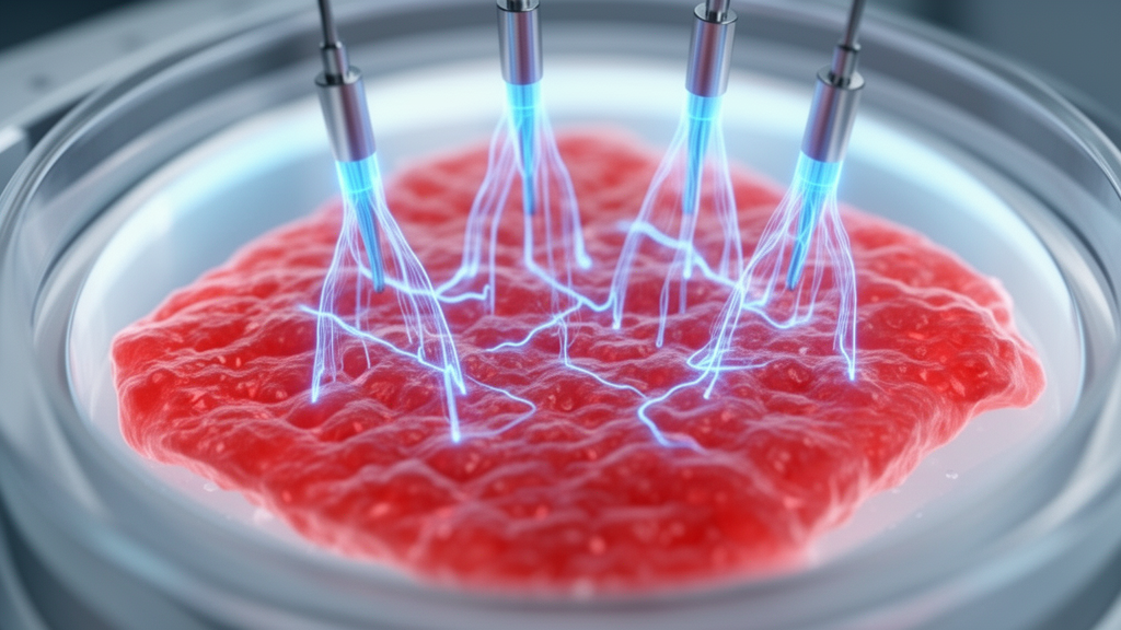
Exogenous electrical stimulation can modulate cell behavior and promote tissue repair across both excitable and non-excitable physiological systems, but translation remains constrained by inconsistent reporting and inadequate electrochemical safety evaluation, according to a mini-review published in Frontiers in Bioengineering and Biotechnology. Author Aaron Lee from the Department of Bioengineering at Imperial College London evaluated findings across in vitro and in vivo studies to examine the coupled relationship linking stimulation paradigms, electrode-electrolyte interface dynamics, and biological outcomes. Electric fields trigger direct electrophysiological responses by altering ionic gradients, depolarizing cell membranes, and activating voltage-gated ion channels, with downstream consequences for gene transcription and translation. However, the review demonstrates that cellular changes reported in the literature often risk conflation with indirect electrochemical alterations in the microenvironment, including localized shifts in pH, metal dissolution, and reactive oxygen species generation.
Biological tissues depend on endogenous bioelectricity to establish homeostatic regulation and govern developmental patterning, morphogenesis, and wound healing. During natural epithelial barrier disruption, local electric field gradients spanning forty to two hundred millivolts per millimeter generate endogenous electric currents between five and ten microamperes per square centimeter to direct epithelial cell migration toward wound margins. In laboratory settings, applying controlled exogenous electric fields serves as a physical guidance cue to steer cellular orientation, elongation, and migration. In integumentary models, human dermal fibroblasts subjected to direct current field strengths between fifty and four hundred millivolts per millimeter exhibit directed electrotaxis toward the anode across exposure periods of five hours, while skin fibroblasts exposed to two hundred millivolts per millimeter demonstrate enhanced transdifferentiation marked by elevated expression of RhoA and Rock1.
For intrinsically excitable tissues, electrical fields reproduce critical physiological dynamics that static culture environments fail to supply. Human pluripotent stem cell-derived cardiomyocytes stimulated with one-millisecond biphasic pulses at field strengths of three to four volts per centimeter show enhanced myofibril organization, accelerated conduction velocities, and improved calcium handling when exposed to seven days of continuous pacing followed by variable frequency regimes. Similarly, murine cardiomyocytes treated with monophasic pulses of one volt at ten hertz for six days exhibit shortened action potential durations to fifty and ninety percent depolarization, improved cellular alignment, and upregulated expression of connexin 43, Kcnh2, and Kcnd2. In musculoskeletal systems, electrical stimulation promotes myogenic and osteogenic commitment, with rat skeletal myoblasts showing increased growth and alpha-smooth muscle actin under 0.15 to 0.3 volts per millimeter fields, and canine myoglossus satellite cells upregulating Pax7, MyoD, myogenin, and myosin heavy chain when pulsed at one hundred and sixty hertz.
Stimulation regimes also exert profound regulatory control over non-excitable support cells and immune populations that orchestrate regenerative niches. Murine bone marrow-derived macrophages exposed to current-controlled pulses of two hundred microamperes at two hertz for four hours exhibit marked reductions in interleukin-1-beta, interleukin-6, and tumor necrosis factor-alpha expression alongside suppressed reactive oxygen species generation, while accumulating intermediate metabolites including citrate, cis-aconitate, isocitrate, and alpha-ketoglutarate. In human monocyte-derived macrophages, voltage-controlled biphasic stimulation at plus-or-minus 2.5 volts and one hertz drives significant migration and upregulates transforming growth factor-beta-one, vascular endothelial growth factor, platelet-derived growth factor-B, and matrix metalloproteinase-9, while suppressing inflammatory cytokine secretion when challenged with lipopolysaccharide.
Neural engineering and nerve repair models similarly reflect the versatility of electrical interventions across structural scales. Neural progenitor cells transplanted into nickel-chromium and polypyrrole nerve guides demonstrate improved functional recovery after sciatic nerve transection, producing increased gastrocnemius muscle mass, elevated compound action potential amplitudes, and restored grip strength. In female Sprague-Dawley rats subjected to contusive spinal cord injury at the L1 and L2 vertebrae, long-term biphasic microstimulation delivered through sputtered iridium oxide film arrays at five microamperes and an estimated field strength of 0.8 to 4.8 millivolts per millimeter improved locomotor recovery on the Basso, Beattie, and Bresnahan scale while reducing response latency.
Despite these phenotypic improvements, the review identifies critical discrepancies in how stimulation is delivered, measured, and reported across the literature. A systematic analysis of experimental parameters reveals pervasive omissions, designated as not reported across published studies for basic physical quantities including charge per phase, charge density per phase, electrode area, and stimulator control mode. Voltage-controlled stimulation is widely implemented in experimental systems due to low circuit complexity and power efficiency, yet it remains fundamentally vulnerable to shifting interfacial charge transfer resistance and variable double-layer capacitance. As a result, identical applied voltages produce drifting, unpredictable currents and disparate electrical fields between preparations. Current-controlled stimulation, theoretical analyses confirm, provides substantially superior reproducibility by maintaining stable charge delivery independent of changing interface impedance, provided stimulator compliance limits are not exceeded.
A primary mechanistic concern highlighted by the author is the widespread operation of stimulation electrodes at potentials that exceed the electrochemical water window. In aqueous biological media, electrolysis begins when electrode polarization surpasses safe thresholds, which nominally span from minus 0.6 to plus 0.8 volts versus silver/silver chloride for platinum electrodes, triggering hydrogen and oxygen evolution. When high overpotentials are imposed, Faradaic reactions govern the interface, generating chemical byproducts including bioactive hydrogen peroxide, corrosive metal ions, and pronounced local pH gradients. In experiments using stainless steel wires or unbuffered platforms, anodic polarization can induce severe pitting corrosion and loss of passivation, introducing toxic oxidation products directly into culture chambers or living tissues. In several reported studies, biological outcomes attributed exclusively to direct bioelectric field sensing may instead reflect cellular adaptation or compensatory antioxidant responses provoked by electrochemically generated chemical species.
To overcome these translational hurdles, future bioelectronic platforms must integrate comprehensive electrochemical characterization and advanced material designs. The review recommends routine pre-experimental assessment using electrochemical impedance spectroscopy, cyclic voltammetry, and voltage transient analysis to measure interfacial impedance, quantify real electrochemical surface area, and delineate the capacitive and Faradaic operational regimes. Physical modifications that increase electrode nanoscale roughness enlarge charge injection capacity, allowing higher currents to pass through purely capacitive dipole reorientation without provoking destructive electron transfer. Furthermore, matching working and counter electrode compositions, employing high-surface-area platinum counter electrodes, and utilizing charge-balanced biphasic waveforms prevent progressive electrode polarization and suppress cumulative charge accumulation.
The engineering of dynamic biological interfaces offers promising avenues to couple bioelectric fields with biosimilar, compliant architectures. Furthermore, scaffold functionalization with piezoelectric, triboelectric, or magnetoelectric materials enables non-invasive cell actuation via external ultrasound or magnetic fields without requiring tethered wired leads. Innovative metabolic energy harvesting systems, such as catalytic electrodes driving the enzymatic oxidation of glucose to gluconate, demonstrate that stable bioelectric currents can be generated continuously from endogenous physiological metabolites to power regenerative devices in situ without standard batteries.
Long-term clinical translation will ultimately require transitioning from static open-loop stimulation protocols to adaptive, closed-loop bioelectronic systems. In vivo, multicellular complexity introduces anisotropic field propagation and risks off-target recruitment of sensitive neighboring nerve fibers, which can induce sensation or adverse pain. Integrating finite element computational modeling with continuous impedance monitoring will allow researchers to predict field steering, maintain precise charge dosing within established Shannon safety limits, and adjust waveforms in real time as the regenerative microenvironment matures.
Subject of Research: Biomedical engineering review evaluating electrical stimulation mechanisms, electrochemical interface safety limits, and reporting standards in tissue repair
Article Title: Sparking regeneration with electrical stimulation for enhanced tissue engineering and repair
Article References: Lee, A. (2026). Sparking regeneration with electrical stimulation for enhanced tissue engineering and repair. Frontiers in Bioengineering and Biotechnology, 14, 1952737. https://doi.org/10.3389/fbioe.2026.1952737
Image Credits: AI Generated
DOI: 10.3389/fbioe.2026.1952737
Keywords: electrical stimulation, tissue engineering, regenerative medicine, bioelectronics, electrophysiology, Faradaic reactions, capacitive charge transfer, electrochemical safety, water window, cellular electrotaxis, cardiomyocyte maturation, neural repair, macrophage modulation, conductive scaffolds, charge density
News Source: Gregory Coleman. (November 20, 2026). Bioelectric Stimulation Shows Tissue Engineering Promise but Requires Rigorous Interface Safety Standards. Scienmag.