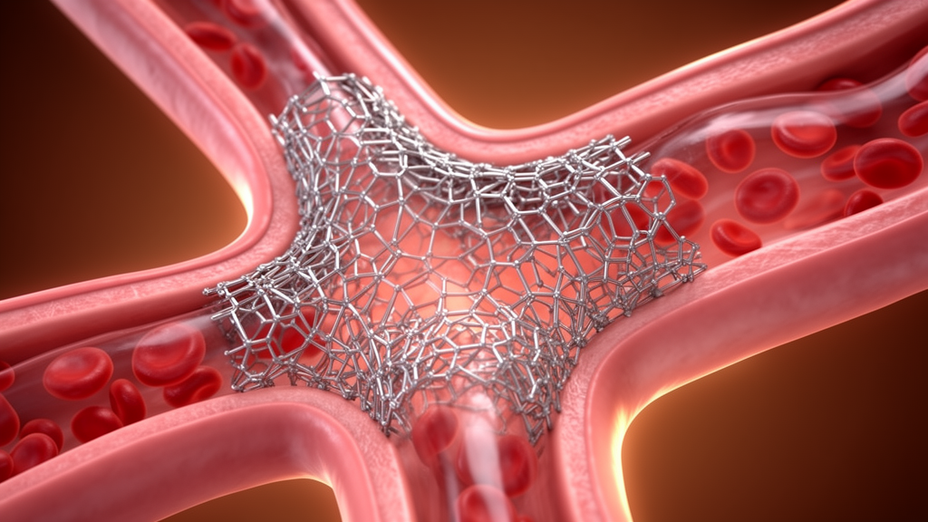
In an interventional cardiology feasibility study evaluating novel dynamic vessel scaffolds, clinicians at Hanoi Heart Hospital successfully performed complex bifurcation percutaneous coronary interventions using the culotte stenting technique with the DynamX bioadaptor implant. The single-center prospective investigation enrolled thirteen consecutive patients presenting with true coronary bifurcation disease between February and October 2025. Evaluating acute mechanical performance and geometry through comprehensive intravascular ultrasound imaging, the research team demonstrated one hundred percent technical and procedural success rates across all treated lesions. The bioadaptor platform, engineered with uncaging helical strands designed to unlock months after deployment, achieved complete device delivery, systematic distal rewiring through deployed struts, and satisfactory acute vessel expansion without any observed strut fractures or apposition failures. These findings offer the first systematic clinical evidence confirming that the unique mechanical properties of this dynamic coronary implant can withstand the severe deformation and overlapping metal architecture inherent to two-stent bifurcation reconstructions.
Coronary artery bifurcations represent approximately fifteen to twenty percent of all percutaneous coronary interventions and remain widely recognized among interventional cardiologists as technically challenging anatomical subsets. Despite continuous technological advances in contemporary drug-eluting stent designs and procedural protocols, bifurcation interventions continue to encounter higher frequencies of adverse procedural complications, target lesion restenosis, and stent thrombosis compared to straightforward non-bifurcation lesions. While interventional guidelines generally advocate a provisional single-stent approach as the primary strategy for most bifurcation lesions, complex two-stent techniques are deemed necessary in twenty to thirty percent of true bifurcations. Such advanced reconstructions are particularly warranted when the side branch vessel exhibits significant caliber, carries diffuse disease extending beyond the ostium, or supplies a substantial territory of viable downstream myocardium that cannot be compromised.
Among established two-stent bifurcation techniques, the culotte approach provides thorough mechanical coverage of both the carinal ridge and the side branch ostium. When paired with systematic proximal optimization techniques and final kissing balloon inflations, culotte stenting yields favorable anatomical outcomes. However, the procedure imposes rigorous mechanical demands upon the chosen implant platform. Favorable execution requires ultra-thin strut architecture beneath eighty micrometers to curb cumulative metal burden and local hemodynamic disturbance, sufficient flexibility to traverse angulated anatomical pathways, robust radial strength to resist recoil during high-pressure balloon expansions, and wide expansion margins capable of accommodating proximal-to-distal vessel caliber discrepancies without structural disruption. Most critically, culotte reconstruction requires flawless guidewire recrossing through implanted stent cells to enable sequential lumen enlargements, making stent cell geometry a pivotal determinant of procedural success.
The investigated DynamX bioadaptor, manufactured by Elixir Medical Corporation, introduces a distinct biomechanical concept compared to conventional permanent drug-eluting metallic stents. Fabricated from an L605 cobalt-chromium alloy with ultra-thin seventy-one-micrometer struts, the implant features three distinct helical strands interconnected by uncaging elements coated with a bioresorbable polymer. Over approximately six months following implantation, this proprietary polymer coating degrades, permitting the helical metallic strands to disengage and separate. This programmed structural release transitions the device from providing rigid, drug-eluting radial support during initial arterial healing into offering compliant mechanical scaffolding that restores natural vessel compliance, cyclical pulsatility, and adaptive vascular remodeling, whereas conventional metal cages permanently constrain physiological vessel dynamics.
The rationale for examining the bioadaptor in bifurcations builds directly upon favorable clinical evidence documented in earlier randomized trials involving less complex coronary lesions. In the primary BIOADAPTOR randomized controlled trial, the bioadaptor demonstrated statistical noninferiority to standard contemporary zotarolimus-eluting stents at twelve months while displaying superior imaging benchmarks, such as reduced late lumen loss and the reestablishment of pulsatile motion. Extended two-year follow-up revealed a statistically significant sixty-five percent decrease in target lesion failure, registering at 1.8 percent versus 5.5 percent with conventional drug-eluting stents. Furthermore, the multicenter INFINITY-SWEDEHEART randomized controlled trial confirmed sustained clinical efficacy, showing a forty-eight percent reduction in target lesion failure between six months and two years. Preliminary application of double bioadaptors in a culotte configuration had been reported in the exploratory ADAPT-CULOTTE case study, yet systematic verification in a prospective patient cohort remained unaddressed until this analysis.
Conducted in strict adherence to the Declaration of Helsinki under formal approval from the Hanoi Heart Hospital Ethics Committee, the single-center study enrolled patients who gave written informed consent. Eligible participants presented with symptomatic coronary artery disease, objective revascularization indications, and de novo true bifurcation lesions categorized under the Medina classification as 1,1,1, 1,0,1, or 0,1,1 configurations. Anatomical entry requirements mandated a side branch reference vessel diameter of at least 2.5 millimeters by visual estimation, an ostial lesion or disease length exceeding 5 millimeters in the side branch, and the absence of extreme caliber mismatch between the main vessel and the branch. Patients presenting with cardiogenic shock, lesions exhibiting heavy calcification necessitating rotational atherectomy, dual antiplatelet therapy contraindications, or left main coronary artery bifurcations were explicitly excluded from the study protocol.
The study cohort comprised thirteen patients with a mean age of 60.3 years, of whom eleven were male and two were female. Clinical presentations included chronic coronary syndromes in eight patients, non-ST-segment elevation myocardial infarction in three patients, and unstable angina in one patient. Baseline cardiovascular risk factors were prevalent, with nine current or former smokers, five individuals diagnosed with diabetes mellitus, and six individuals receiving treatment for hypertension. Anatomically, target bifurcation interventions were located in the left anterior descending artery and diagonal branch junction in nine patients, the left circumflex and obtuse marginal junction in three patients, and the posterior descending and posterolateral ventricular branch in one patient. Medina 1,1,1 morphology predominated, accounting for twelve of the thirteen treated lesions, while the mean bifurcation angle measured sixty-four degrees.
All surgical procedures were executed via transradial or transfemoral vascular access using six-French or seven-French guide catheters according to an exhaustive ten-step culotte protocol. Operators performed dual-wire lesion preparation with aggressive balloon predilatation and baseline intravascular ultrasound, followed by deployment of the initial bioadaptor sized one-to-one to the distal reference diameter into the more angulated branch, leaving two to three millimeters protruding into the main stem. Following an initial proximal optimization technique expansion, operators rewired the untargeted branch through the deployed strut cells, dilated the crossing cell with small noncompliant balloons, and deployed the second bioadaptor within the main vessel. A second proximal optimization inflation was followed by recrossing into the initial branch, executing a simultaneous final kissing balloon inflation with noncompliant balloons, completing a final proximal optimization shaping, and performing multi-vessel intravascular ultrasound pullbacks.
Procedural execution demonstrated remarkable mechanical compliance, achieving one hundred percent technical success. Across twenty-six distinct rewiring maneuvers required during the two-stent reconstructions, guidewire crossing through bioadaptor strut cells succeeded on the initial attempt in every instance without necessitating compromise into suboptimal proximal positions. Operators successfully achieved intentional distal cell crossing in all twenty-six instances, noting that the helical geometry and uncaging elements provided an accommodating cell configuration for wire navigation. Initial rewiring required a mean duration of 39.2 seconds, whereas subsequent recrossing through double-layered metal required a mean of 206.9 seconds. Complete simultaneous final kissing balloon inflation was achieved in all thirteen patients without persistent balloon waist indentation, with an overall mean procedural time of 55.2 minutes, fluoroscopy time of 38.4 minutes, and contrast volume of 218 milliliters.
High-resolution quantitative intravascular ultrasound imaging conducted with the Boston Scientific AVVIGO guidance system validated robust acute mechanical expansion throughout all treated bifurcations. In the main branch, mean reference vessel area measured 7.21 square millimeters, achieving a mean distal stent area of 7.30 square millimeters and an overall expansion of 90.9 percent. At the main branch ostium, achieved stent area averaged 6.83 square millimeters, representing 94.9 percent of the expected ostial dimension, with 84.6 percent of lesions exceeding eighty percent expansion. Side branch assessments demonstrated a mean reference area of 5.94 square millimeters, a distal stent area of 6.18 square millimeters, an overall expansion of 94.5 percent, and an ostial expansion of 90.2 percent of expected dimensions, while the proximal overlapping main stem attained an expansion ratio of 0.95 without structural collapse.
Qualitative ultrasound evaluation verified comprehensive anatomical coverage across the carinal bifurcation and side branch ostia in every patient, clearly resolving the expected double-layer metallic architecture within the proximal parent vessel. Importantly, imaging confirmed preserved inter-strut spacing and complete vessel wall apposition without any instances of strut fracture, structural deformation, or malapposition exceeding two hundred micrometers. The uncaging elements and protective polymer coatings remained fully intact following aggressive balloon inflations. In isolated side branch ostia exhibiting lower expansion values, suboptimal widening was linked to localized resistance from calcified nodules situated opposite the carina rather than device failure, highlighting the crucial role of imaging guidance during complex bifurcation reconstruction.
In-hospital clinical monitoring confirmed an absence of major adverse cardiac events, with no recorded patient deaths, emergency target vessel revascularizations, or acute vessel closures prior to discharge. One patient developed an access-site hematoma that resolved under conservative management. While four patients demonstrated postprocedural high-sensitivity troponin T elevations exceeding five times the normal institutional reference threshold, none exhibited clinical ischemic symptoms, novel electrocardiographic abnormalities, or angiographic flow impairment. Because preprocedural troponin concentrations were dynamically fluctuating or lacked stable baselines, these biomarker shifts could not be definitively attributed to intraprocedural necrosis, resulting in zero adjudicated periprocedural myocardial infarctions and a cumulative procedural success rate of one hundred percent.
The acute findings provide encouraging support for utilizing dynamic bioadaptor technology within complex bifurcation scenarios, where permanent metal cages frequently impair long-term shear stress patterns and provoke accelerated neoatherosclerosis. Although double metal layers proximally cannot be avoided during culotte stenting, the anticipated degradation of the polymer matrix at six months allows independent strand separation, potentially diminishing the rigid hemodynamic footprint of conventional stents while facilitating adaptive vascular remodeling. The investigators underscored several study limitations, including the small cohort size of thirteen patients, the single-center setting involving highly experienced operators, the lack of a randomized comparator arm, and the restriction to acute intravascular imaging. Future multicenter investigations incorporating optical coherence tomography and long-term clinical surveillance will be required to confirm whether bioadaptor uncaging translates into durable physiological superiority in complex bifurcations.
Subject of Research: Feasibility and intravascular ultrasound outcomes of DynamX bioadaptor implantation in culotte coronary bifurcation stenting
Article Title: Feasibility and short-term outcomes of bioadaptor in bifurcation intervention using culotte technique: intravascular ultrasound analysis from a single-center experience
Article References: Hoang, V., Tran, G. T., Ngoc, D. T., Dang, D. N., Van, D. T., Hung, C. P., & Dinh, T. T. (2026). Feasibility and short-term outcomes of bioadaptor in bifurcation intervention using culotte technique: intravascular ultrasound analysis from a single-center experience. Journal of Cardiovascular Imaging, 34(1), Article 20. https://doi.org/10.1186/s44348-026-00083-8
Image Credits: AI Generated
DOI: 10.1186/s44348-026-00083-8
Keywords: DynamX bioadaptor, culotte stenting, coronary bifurcation, percutaneous coronary intervention, intravascular ultrasound, interventional cardiology, bifurcation lesions, stent expansion, stent apposition, distal cell crossing, kissing balloon inflation, proximal optimization technique, coronary artery disease, cardiovascular imaging, target lesion failure, uncaging implant, Hanoi Heart Hospital
News Source: Ophelia Keating. (November 18, 2026). Bioadaptor Culotte Stenting Shows Technical Feasibility in Complex Coronary Bifurcations. Scienmag.