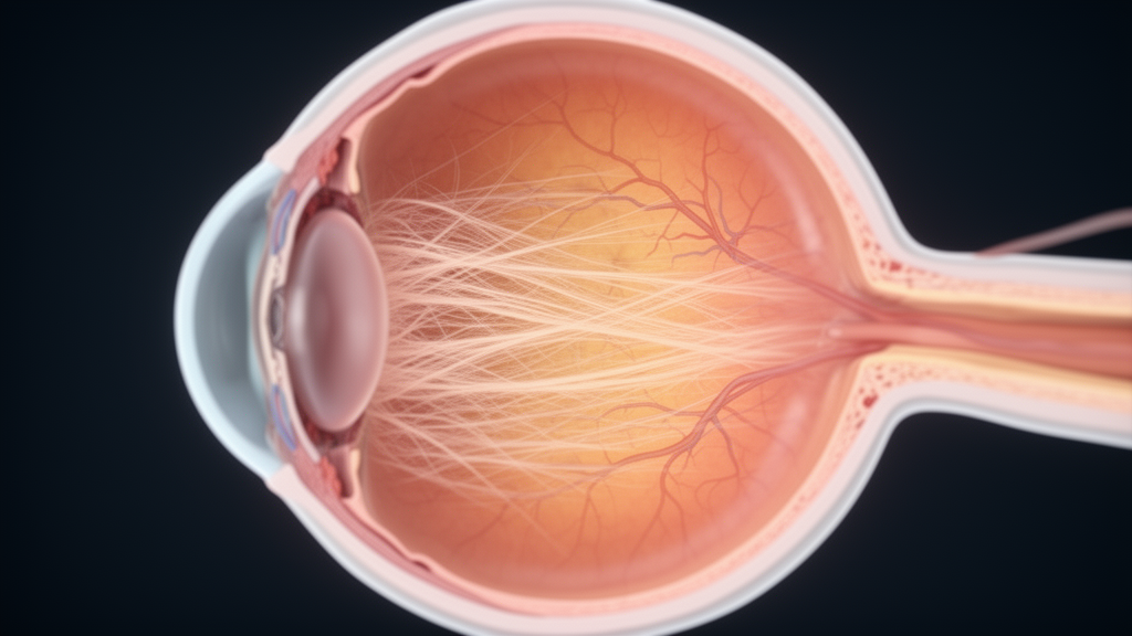
Two teenagers with an unusual pattern of retinal scarring are at the centre of a new case report that challenges the usual way ophthalmologists think about peripapillary subretinal fibrosis, a condition in which scar-like fibrotic tissue forms beneath the retina near the optic nerve head. The report, published in BMC Ophthalmology by Selda Celik Dulger and Mehmet Yasin Teke, describes two adolescent patients who developed bilateral peripapillary subretinal fibrosis without any overt clinical or angiographic evidence of active inflammation inside the eye. Because the condition has almost always been linked to inflammatory eye disease, the authors deliberately describe their findings as a “presumed non-inflammatory phenotype,” a phrase chosen to signal that the label is a clinical description rather than a proven diagnosis.
Peripapillary subretinal fibrosis is an uncommon retinal finding that has most frequently been reported in association with inflammatory chorioretinal disorders, particularly multifocal choroiditis and panuveitis. In those diseases, fibrosis tends to develop at sites of previous inflammatory lesions and can spread toward the peripapillary region or the macula, the central part of the retina responsible for sharp vision, causing permanent structural damage and irreversible visual loss. The scarring has also been documented in inherited retinal disorders, most notably in patients with biallelic mutations in the NR2E3 gene, where fibrosis occurs as part of a broader degenerative spectrum that includes outer retinal atrophy, the nummular deposits characteristic of enhanced S-cone syndrome, and helicoid subretinal lesions. Against that background, bilateral fibrosis in young patients with no visible signs of inflammation is a genuinely puzzling presentation.
The study was a retrospective observational case series based on clinical records and multimodal imaging of two adolescent patients evaluated at a tertiary referral centre. Both patients were followed for approximately four years with serial clinical examinations and imaging that included colour fundus photography, fluorescein angiography, and spectral-domain optical coherence tomography. Because the cases were historical, the available records were incomplete: the results of genetic testing, electrophysiological evaluation, and systemic laboratory investigations, if they were performed during the original work-up, could not be verified. The authors state plainly that inherited retinal disease and subclinical inflammatory or infectious disease therefore could not be definitively excluded, a caveat that shapes every conclusion in the paper.
The first patient was a 14-year-old male whose best-corrected visual acuity was 6/7.5 in the right eye and counting fingers at 5 metres in the left eye. Anterior segment examination was unremarkable, with no cells or haze, and fundus examination revealed bilateral peripapillary subretinal fibrosis. In the left eye the fibrotic lesion extended from the peripapillary region toward the macula and involved the foveal area, while the right eye showed a lesion confined to the peripapillary region. No multifocal chorioretinal scars, punched-out lesions, or signs of active inflammation were observed, and areas of hyperpigmentation were present within the fibrotic region.
Fluorescein angiography in this patient demonstrated late staining of the fibrotic areas, while the hyperpigmented regions blocked fluorescence. In the macula of the left eye, a hyperfluorescent lesion surrounded by a hypofluorescent halo was seen, a pattern suggestive of a subfoveal choroidal neovascular lesion. Because OCT angiography was not available from the original evaluation, the vascular lesion could not be further classified with that technique. Spectral-domain optical coherence tomography of the right eye showed a subretinal hyperreflective lesion temporal to the optic disc with preserved macular architecture, while the left eye had two dome-shaped hyperreflective subretinal lesions, one temporal to the disc and one beneath the fovea, along with small intraretinal cystic spaces suggestive of choroidal neovascularisation and outer retinal tubulation in the peripapillary region. Two intravitreal anti-vascular endothelial growth factor injections were administered to the left eye, although the historical records did not allow reliable verification of pre- or post-treatment visual acuity, OCT response, injection interval, neovascular subtype, or recurrence data.
The second patient was a 16-year-old female with best-corrected visual acuity of 6/6 in the right eye and counting fingers at 3 metres in the left eye. Slit-lamp examination revealed no signs of anterior segment inflammation. Fundus examination again demonstrated bilateral peripapillary subretinal fibrosis, with a fibrotic extension in the left eye that reached and involved the macula, where marked macular atrophy was present, while the right eye had a peripapillary lesion without macular involvement. No additional chorioretinal lesions or vitreous haze were identified. Fluorescein angiography showed late staining of the fibrotic areas consistent with scar tissue, and hyperfluorescence in the left macula due to a window defect, without evidence of leakage. Optical coherence tomography of the right eye showed subretinal hyperreflective scar tissue temporal and nasal to the optic disc with a preserved foveal contour and outer retinal layers, whereas the left eye showed macular atrophy involving the outer retinal layers beneath the fovea, associated subretinal fibrotic tissue, and increased choroidal hypertransmission secondary to retinal pigment epithelium atrophy.
One of the most clinically informative observations in the series is the relationship between anatomy and vision. Visual impairment was observed exclusively in eyes with macular extension of the fibrosis, whereas eyes whose lesions remained confined to the peripapillary region preserved normal visual acuity. The authors conclude that visual prognosis in this presentation appears to be determined primarily by macular involvement rather than by the presence of peripapillary fibrosis itself. During the approximately four-year follow-up, no clinically apparent enlargement of the lesions, development of new lesions, or progression toward the macula was documented in the available records, suggesting a relatively stable course, although the authors note this stability is based only on what the records captured.
The discussion grapples honestly with what the findings might mean. The consistent peripapillary localisation, bilateral involvement, and absence of accompanying inflammatory signs distinguish these cases clinically from the classical presentation of multifocal choroiditis and related inflammatory disorders. However, the authors emphasise that the absence of overt inflammatory findings cannot exclude previous or subclinical inflammation, which is why they frame the cases as a possible or presumed non-inflammatory phenotype rather than a definitively established disease entity. In inflammatory chorioretinal disorders, subretinal fibrosis is thought to arise from chronic or recurrent damage to the outer retina, retinal pigment epithelium, and choroid, with subsequent activation of fibroblasts, myofibroblasts, and retinal pigment epithelium-derived cells leading to extracellular matrix deposition and scar formation. The absence of clinical or angiographic evidence of inflammation in these two patients suggests that alternative mechanisms may be involved, and the authors propose possibilities including primary retinal pigment epithelium dysfunction, localised microvascular insufficiency, or developmental anomalies affecting the peripapillary region.
The inherited disease possibility receives particular attention because of the patients’ age. Peripapillary subretinal fibrosis has been described in association with biallelic NR2E3 mutations, where it may occur alongside outer retinal atrophy and characteristic pigmentary changes. Although no characteristic clinical or multimodal imaging features suggestive of NR2E3-related retinopathy were identified in these cases, the authors state that an underlying inherited retinal disorder could not be definitively excluded in the absence of verifiable genetic and electrophysiological testing. They accordingly recommend that the differential diagnosis include multifocal choroiditis and panuveitis, NR2E3-associated retinal disease, serpiginous choroiditis, and other inherited retinal disorders characterised by outer retinal degeneration, retinal pigment epithelium abnormalities, macular atrophy, or helicoid and peripapillary lesions. In the first patient, the suspected subfoveal choroidal neovascularisation also raises the question of whether the fibrotic changes are causally related to neovascular processes; the authors note that the temporal relationship remains uncertain and that, given the stable bilateral peripapillary distribution and absence of inflammatory signs, the suspected neovascularisation may represent a secondary complication.
The authors suggest that recognising this pattern may help clinicians avoid prematurely classifying such findings as inflammatory chorioretinal disease, potentially reducing unnecessary investigations or inappropriate immunosuppressive treatment. At the same time, the limitations are considerable: the series contains only two patients, and the inability to verify historical genetic, electrophysiological, systemic laboratory, and OCT angiography investigations means the underlying cause remains open. The morphological variability between the two patients may reflect different stages of a common underlying process, differences in chronicity, or heterogeneity in pathogenic mechanisms, and the authors caution that the cases may represent different manifestations within a broader spectrum of bilateral peripapillary subretinal fibrosis rather than a single established clinical entity. Further studies, including genetic and electrophysiological evaluation, are required to better define the mechanisms behind this presentation, and until such data exist, the report stands as a carefully documented description of a rare and diagnostically challenging pattern rather than the establishment of a new disease.
Subject of Research: A retrospective case series of two adolescents with bilateral peripapillary subretinal fibrosis lacking overt clinical or angiographic evidence of active intraocular inflammation.
Article Title: Presumed non-inflammatory bilateral peripapillary subretinal fibrosis in adolescent patients
Article References: Celik Dulger, S., & Teke, M. Y. (2026). Presumed non-inflammatory bilateral peripapillary subretinal fibrosis in adolescent patients. BMC Ophthalmology, 26(1), Article 686. https://doi.org/10.1186/s12886-026-05466-5
Image Credits: AI Generated
DOI: 10.1186/s12886-026-05466-5
Keywords: peripapillary subretinal fibrosis, retina, adolescents, case report, multifocal choroiditis, NR2E3, optical coherence tomography, fluorescein angiography, choroidal neovascularisation, macular atrophy, ophthalmology, BMC Ophthalmology
News Source: Ophelia Keating. (October 29, 2026). Bilateral Retinal Fibrosis Without Inflammation Reported in Two Teenagers. Scienmag.