A little-known population of intestinal epithelial cells may help explain how the gut balances fluid, electrolytes, acidity and mucus—and how that system can become disrupted during diarrhea. Known as BEST4⁺ cells, the cells have emerged from single-cell and spatial transcriptomic studies of human, pig, rat and other vertebrate intestines. A review by Hao-zhan Qu and Xiu-qi Wang presents them as a possible cellular hub connecting normal ion transport with secretory diarrhea. The authors emphasize, however, that much of the evidence remains indirect or comes from organoids and comparative transcriptomic analyses. Whether BEST4⁺ cells are direct viral targets, or instead respond to signals released by infected cells, remains unresolved.
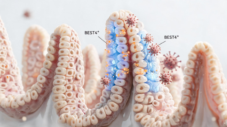
BEST4⁺ cells are defined by a distinctive combination of genes, including BEST4, OTOP2, CA7 and GUCY2C. In the small intestine, they also show particularly high expression of CFTR, the chloride and bicarbonate channel best known for its role in cystic fibrosis. Together, these markers suggest a specialized role in moving negatively charged ions across the epithelium. Chloride and bicarbonate transport draw water into or out of the intestinal lumen and help regulate the chemical environment surrounding epithelial cells and mucus. OTOP2 adds a sensing capability: this proton-selective channel can respond to changes in extracellular acidity. CA7 may support intracellular bicarbonate production, while GUCY2C encodes guanylyl cyclase C, a receptor that converts extracellular signals into cyclic GMP. The resulting molecular profile is unusually coherent for a relatively small cell population.
The cells were first associated with the intestinal epithelium through studies of bestrophin expression, but modern single-cell sequencing made their identity much clearer. These analyses separate individual epithelial cells according to their RNA profiles, allowing researchers to identify populations that conventional staining can overlook. BEST4⁺ cells appear early in human intestinal development, reportedly as early as gestational week 11, and generally represent less than 5 percent of the fetal epithelial compartment. In adults, their abundance varies by region, with reported enrichment in parts of the jejunum, ileum and colon. Small-intestinal cells are concentrated toward the upper and middle villus, whereas colonic cells tend to occupy apical crypt regions. The regional differences suggest that a shared core program may be adapted to local pH, microbial exposure, mucus and transport demands.
The developmental identity of BEST4⁺ cells is still being debated. Several lines of evidence place them near the end of an absorptive differentiation pathway. They occupy post-mitotic compartments, lack conventional proliferation markers and express genes associated with mature enterocytes and colonocytes, including VIL1, AQP8 and SLC26A3. Human organoid experiments indicate that NOTCH signaling is required for their emergence, and the transcription factor SPIB appears indispensable: removing SPIB with CRISPR-based gene editing prevented BEST4⁺ cell generation even when NOTCH signaling remained active. Yet other findings point toward connections with the secretory lineage. Trajectory analyses in human tissue have identified a low-probability link to ATOH1-positive secretory progenitors, while zebrafish lineage-tracing experiments suggest that related cells can arise from secretory precursors. These differences may reflect species, anatomical region or inflammatory state rather than a single universal developmental route.
Under normal conditions, BEST4⁺ cells may act as coordinated pH and ion-transport units. BEST4 belongs to the bestrophin family of calcium-activated anion channels, which can conduct chloride and bicarbonate when intracellular calcium rises. CFTR provides another major route for apical anion secretion. The cells also express guanylin and uroguanylin, the endogenous ligands for GUCY2C, alongside the receptor itself. This arrangement could create an autocrine circuit in which locally produced ligands stimulate cyclic GMP, activate downstream protein kinases and increase CFTR activity. Bicarbonate secretion is important beyond fluid balance: it helps neutralize acidity near the epithelial surface and allows newly released MUC2 mucin to hydrate and expand into an effective protective layer. The review therefore proposes that BEST4⁺ cells may support mucus-barrier assembly in cooperation with goblet cells, although direct proof that these cells provide the critical bicarbonate flux is still lacking.
The same machinery can be exploited during secretory diarrhea. Bacterial heat-stable enterotoxin, or STa, binds and activates GUCY2C, raising intracellular cyclic GMP and stimulating CFTR through protein kinase G. Cholera toxin and the heat-labile toxin of enterotoxigenic Escherichia coli activate adenylate cyclase through persistent cyclic AMP signaling, leading to protein kinase A-mediated CFTR activation. In both cases, excessive chloride and bicarbonate secretion promotes water movement into the lumen. The review highlights evidence that BEST4⁺ cells are unusually equipped for this response because they co-express GUCY2C and CFTR at functionally relevant levels. Human intestinal organoids exposed to these pathways swell as fluid accumulates. Investigational inhibitors of GUCY2C or CFTR can reduce secretion in experimental systems, but broad suppression carries risks because basal GUCY2C signaling also contributes to barrier integrity, mucus hydration and epithelial maintenance.
Viral diarrhea may involve BEST4⁺ cells more indirectly. Porcine epidemic diarrhea virus preferentially damages villus absorptive enterocytes and can impair NHE3, a sodium-hydrogen exchanger needed for sodium-coupled water absorption. Studies in infected piglets have reported reduced expression of several water and nutrient transporters, together with increased ileal CFTR transcripts. Electrical measurements of infected jejunal tissue also indicate enhanced secretory responses. These observations are consistent with a shift away from absorption and toward secretion, and the presence of CFTR-rich BEST4⁺ cells makes them plausible contributors. But the available evidence does not show that the virus infects BEST4⁺ cells or that the cells are responsible for the altered current. Rotavirus offers another possible route: its NSP4 protein disturbs calcium signaling and triggers ADP-dependent calcium waves in neighboring uninfected cells. BEST4⁺ cells could respond as bystander effectors through calcium-sensitive anion channels, but this remains a testable hypothesis.
Norovirus likewise causes changes that could intersect with the BEST4⁺ program, including reduced epithelial resistance and increased electrogenic chloride secretion. The virus can replicate in differentiated enterocytes and some enteroendocrine cells, but there is no direct evidence that mature BEST4⁺ cells support norovirus replication. Inflammation may nevertheless alter their numbers or activity. In human organoids, interferon-gamma increases BEST4⁺ cell differentiation through a SPIB-dependent mechanism, and the resulting cells show stronger CFTR-dependent secretion after toxin exposure. If antiviral inflammation produces a similar response in living intestine, it could amplify fluid loss during acute disease. Conversely, bicarbonate secretion and mucus hydration might aid barrier repair during recovery. The review also connects BEST4⁺ cells to inflammatory bowel disease, where their abundance and expression of transport and metal-buffering genes appear altered, and to cystic fibrosis, in which defective CFTR trafficking may leave these high-CFTR cells unable to regulate anion transport, luminal acidity and mucus hydration.
These possibilities make BEST4⁺ cells attractive but challenging therapeutic targets. A drug that blocks pathological GUCY2C or CFTR activation could reduce fluid loss, yet permanent or systemic inhibition might undermine normal mucosal defense. Experimental CFTR inhibitors have reduced toxin-induced secretion in rodents, although some show limited solubility, rapid washout, narrow dosing windows or off-target effects on mitochondria. A more selective strategy may involve ADRA2A, an adrenergic receptor enriched in human BEST4⁺ cells; activating it suppresses cyclic AMP secretion and reverses cholera-toxin-induced swelling in enriched organoids. Such findings remain preclinical. The next steps will require lineage-specific genetic tools, direct electrophysiological measurements and disease experiments in animals that actually possess a conserved BEST4⁺ population. Conventional laboratory mice lack a canonical intestinal Best4 lineage, making rats, pigs, zebrafish and human organoids complementary rather than interchangeable models. Until researchers can manipulate these cells in vivo, BEST4⁺ cells should be viewed not as a confirmed master switch for viral diarrhea, but as a promising framework for understanding how infection, inflammation and epithelial ion transport converge.
At the molecular level, the proposed hub function depends on the way several transport systems are colocated rather than on BEST4 alone. Bestrophin channels are described as pentameric calcium-sensitive anion channels with a calcium-binding region, a hydrophobic gate and a cytoplasmic regulatory segment. This architecture provides a potential link between intracellular calcium signals and rapid changes in chloride or bicarbonate permeability. In a BEST4⁺ cell, such calcium-dependent conductance could complement CFTR, whose activity is controlled primarily through cyclic-nucleotide signaling. The two routes therefore offer distinct but potentially convergent means of regulating apical anion movement, while OTOP2 and CA7 could help couple that transport activity to the chemical conditions at the epithelial surface.
That arrangement also helps explain why anatomical location matters. BEST4⁺ cells are reported in the proximal small intestine and at the colonic surface, but their associated transport programs are not identical in every region or species. Villus-associated small-intestinal cells encounter different nutrient, acid and fluid gradients from cells near colonic crypt openings. Cross-species conservation supports a shared cellular program, yet conservation of marker genes does not establish conservation of net ion flux. Differences in epithelial architecture, microbiota, mucus organization and channel abundance could alter the physiological contribution of the same transcriptional cell type. Functional comparisons will therefore need to measure transport in defined regions rather than treating all BEST4⁺ cells as equivalent.
A central experimental challenge is separating correlation from cell-specific causation. High BEST4, GUCY2C or CFTR expression identifies a candidate effector population, but whole-organoid swelling, tissue short-circuit current and bulk transporter measurements integrate responses from many epithelial cells. Stronger tests would combine selective deletion or activation of BEST4⁺ cells with live measurements of intracellular pH, calcium, cyclic GMP, bicarbonate flux and mucus expansion. These experiments could determine whether BEST4 itself is the principal anion pathway, whether it mainly amplifies CFTR-mediated secretion, or whether its greatest contribution is sensing and coordinating responses among neighboring cells. They could also clarify whether toxin-induced secretion requires the endogenous guanylin–uroguanylin circuit or is driven predominantly by pharmacological stimulation of GUCY2C.
Therapeutic development will depend on preserving the distinction between pathological hypersecretion and protective basal transport. GUCY2C signaling and bicarbonate movement may contribute to epithelial maintenance and mucus function even while excessive cyclic GMP or cyclic AMP drives diarrhea. This argues for approaches that limit abnormal signal amplitude, duration or cellular targeting instead of eliminating the pathway entirely. Cell-type-resolved physiology, supported by species with a conserved BEST4⁺ population, should help identify that therapeutic window and establish whether the proposed hub is a druggable controller or primarily a useful map of interacting intestinal transport mechanisms.
Subject of Research: BEST4⁺ intestinal epithelial cells and their role in ion transport and diarrheal mechanisms
Article Title: BEST4⁺ cells: a potential hub of intestinal ion transport and diarrhea manipulation
Article References: Qu, H.-Z., & Wang, X.-Q. (2026). BEST4⁺ cells: a potential hub of intestinal ion transport and diarrhea manipulation. Advanced Biotechnology, 4(3), Article 33. https://doi.org/10.1007/s44307-026-00126-7
Image Credits: AI Generated
DOI: 10.1007/s44307-026-00126-7
Keywords: BEST4⁺ cells, intestinal epithelium, ion transport, CFTR, GC-C signaling, secretory diarrhea, viral gastroenteritis, mucus barrier, BEST4, cells, potential, intestinal
Cite Scienmag News
APA MLA Chicago
Scienmag. (August 28, 2026). BEST4⁺ Intestinal Cells May Link Ion Transport to Viral Diarrhea. https://scienmag.com/best4%e2%81%ba-intestinal-cells-may-link-ion-transport-to-viral-diarrhea/
Scienmag. “BEST4⁺ Intestinal Cells May Link Ion Transport to Viral Diarrhea.” Scienmag, 28 August 2026, https://scienmag.com/best4%e2%81%ba-intestinal-cells-may-link-ion-transport-to-viral-diarrhea/. Accessed 28 August 2026.
Scienmag. “BEST4⁺ Intestinal Cells May Link Ion Transport to Viral Diarrhea.” Scienmag. August 28, 2026. https://scienmag.com/best4%e2%81%ba-intestinal-cells-may-link-ion-transport-to-viral-diarrhea/
Copy citation Download RIS
Tags: BEST4BEST4 positive cells in gutBEST4⁺ cellscellsCFTRchloride and bicarbonate channels in intestineGC-C signalinggut mucus regulation and ion movementintestinalintestinal cell markers and gene expressionintestinal epithelial cell functionintestinal epithelial response to infectionintestinal epitheliumion transportion transport and diarrheamucus barrierpotentialrole of CFTR in intestinal healthsecretory diarrheasecretory diarrhea mechanismssingle-cell transcriptomics intestinal cellsspatial transcriptomics gutviral gastroenteritisviral infection impact on intestinal cells